Tritrichomonas foetus
Location and host
- This parasite is found in prepuce uterus, small intestine of cattle, cat.
- Also occur in pig, horse, deer.
Morphology
- Organism is pear-shaped , approximately 10-25 µm long and 3-15 µm wide
- It has single nucleus and four flagella each arising from a basal body situated at anterior rounded ends
- Three of flagella are free anteriorly, while the fourth extend backwards to form undulating membrane along the length of organism and then continues posterior as a free flagellum
- Axostyle, hyaline rod with skeletal function, extends the length of cell and usually projects posteriorly. Anterior end of axostyle is enlarged to form capitulum.
- Coasta is prominent but there is no pelta
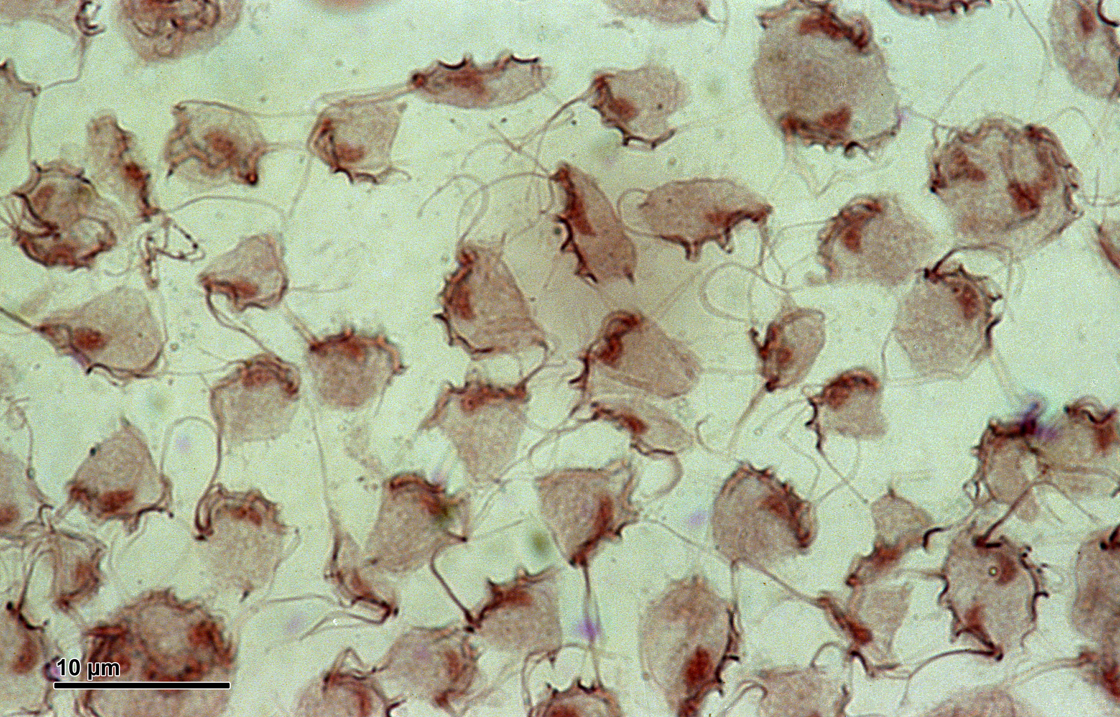
Fig: Tritrichomonas foetus
- In fresh preparations, organisms is motile and progresses by rolling jerky movements
Life cycle
- Transmission occurs through coitus
- When infected bull mates with cow, parasite goes inside the vagina of cow
- There they undergo multiplication by binary fission
- This results in vaginitis by third day, post infection
Transmission
- Transmission occurs through coitus under natural condition
- Artificial insemination. Teaser bulls and gynecological examination may become source of infection, if no proper precautions are taken
- Bulls, once infected, remain permanent source of infection
Pathogenesis
- Disease caused by this organism is called bovine trichomoniasis
In cow
- Organism gets entry into genital cavity of cow through coitus or artificial insemination
- Organism reaches vagina where they multiply primarily in vagina, causing vaginitis by third day post infection
- Organism then invade uterus through cervix and may disappear from vagina or may remain there producing low-grade inflammation and catarrh
- Slight reddening and swelling of vulva and vagina may be seen
- Initial lesion may vary in intensity which may be slight as to pass unnoticed or it may produce a mucopurulent discharge
In bull
- Preputial discharge associated with small nodules on preputial and penile membrane may develop shortly after infection
- Organism are present in preputial cavity of bulls, with come concentration in fornix and around glans penis
Clinical signs
In bulls
- Pain during micturition and disinclination to serve cows
- Mucopurulent discharge may be present
- Small red nodules in the mucous surface of prepuce
- Swelling and pain subside in chronic case. Infected bulls remain permanently infected
In cows
- Characterized by early abortion. Abortion usually occurs 8-16 weeks after conception
- Placentitis with detachment of placental membrane and death of fetus lead to abortion usually between 8-16 weeks after infected service. It is followed by uterine discharge and animals may show a series of irregular heat period.
- Maceration of fetus leads to catarrhal or purulent endometritis and closure of cervix with retained corpus leuteum leads to closed pyometra. This visually stimulates the appearance of pregnancy. Uterus may contain several liters of thin, greyish-white fluid swarming with trichomonads.
- Retention of developing fetal membranes leads to a purulent endometritis, persistent uterine discharge and anestrus.
- In some case, despite infection, pregnancy is not terminated by a abortion and a normal full-term calf is born
- Irregular estrus cycle
- Cow usually recovers and generally becomes immune for that breeding season ( at least), after infection or abortion.
Two types of abortion can be seen
- 1st type: Both the fetus and fetal membrane are expelled out. Damage is less to wall of uterus
- 2nd type: Only fetus comes out but the fetal membrane is retained in uterus. These begins to petrify resulting in pyometra. In this case, cow usually become sterile.
Diagnosis
- Diagnosis is based on herd history, demonstration of organism and serological investigation
- Herd history includes. Signs of early abortion, repeated returns to service or irregular estrus cycle
- Confirmatory diagnosis is through demonstration of organism in placental fluid, stomach contents of aborted fetus, uterine washing, pyometra discharge or vaginal mucus.
- Vaginal mucus or exudate or saline washing from vagina and preputial cavity can be examined microscopically. Samples to be examined should be kept warm throughout by adding warm saline
- In case of pyometra, pus should be examined as it may contain plenty of foetus.
- Since the organism are present intermittently, examination may need to be repeated several times
- Organism can be cultured in vitro in diamond’s medium, clausen’s medium or trichomonas medium
- Serological investigation includes cervical mucus agglutination test and skin test. For cervical mucus agglutination test, mucus is obtained by suction from anterior end of vagina a few days after estrus mucus is mixed with glucose saline and suspension of invitro cultured foetus is used.
- At least three negative test and two normal estrus period can indicate absence of infection in cow
- A bull is considered uninfected when six test at weekly intervals are negative and at least two virgin heifers breed free from infection shows no infection
- Cultural method: Parasite can be cultured in peptone broth media to which 10% cattle serum and antibiotic like penicillin or streptomycin is added. Culture is kept at 370 In positive case, parasite will grow within 28 hrs.
Treatment
In bulls
- Wash the penis with weak solution of detergent, dry it and Flavine ointment is introduced into the preputial cavity and massaged for 15-20 minutes. Then washing with 1% Acriflavine solution to kill the parasite
- Dimetridazole given @ 50 mg/kg body weight, parenterally for 5 days
- Animal should be given breeding rest and AI should be done with clean uninfected semen
In cows
- Wash vagina and uterus with 1% acriflavine or 3% lactic acid solution. Berenil 1% solution 100-150 cc is then put into uterus and keep for 15-30 minutes to kill parasite
- Dimetridazole is given orally or intravenously @ 50 mg/kg body weight for 5 days
Control
- All infected bulls must be castrated or slaughtered
- Bulls should be examined prior to breeding
- AI instruments should be sterilized properly
- Aborted cows should be given breeding rest for three consecutive oestrus periods
- Proper maintenance of records of service dates, calving, etc.