Theileria annulata
Location and host
- These species of parasites are found in blood and lymphatics of cattle and buffalo. Vectot for this parasite is Hyalomma species of tick.
Morphology
- Trophozoites forms in erythrocyte are round to oval; but may also be rod-shaped or comma shaped.
- Division by binary fission may form two or four daughter cells. Four daughter cells appear in shape of cross (Maltese cross).
- Koch bodies are found in lymphocytes of spleen or lymph nodes or even free in these organs. They measure about 8 µm but can be upto 27µm.
- Two types of meronts occur; Macro meronts containing chromatin granules of 0.4-1.9 µm in diameter. They divide further to become micro meronts that contain chromatin granules of 0.3-0.8µm in diameter. They produce merozoites of 0.7-1 µm in diameter.

Lifecycle
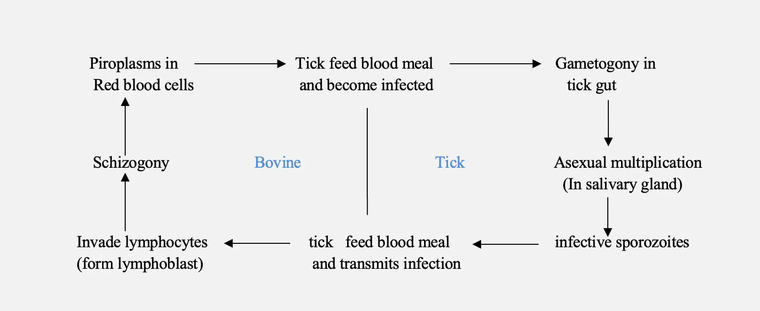
- Under natural condition, susceptible animals are infected by tick vector containing number of sporozoites (uninucleate infective particle) during its attachment for engorgement.
- Transmission does not occur immediately on attachment because sporozoites develop in salivary glands during first 2-4 days of engorgement of nymphal and adult stage of ticks.
Note: Synchronously : recurring or operating at exactly the same periods.
Subsequently: After wards
- Sporozoites released then enter lymphocytes. This stimulate lymphoproliferation and parasite or host cell divide synchronously.
- After several days, parasite appear in lymph node and in lymphoid and reticulo endothelial tissues as multi nucleated macro schizonts called ‘Koch’s bodies’. Each macroschizoints contain about 8 nuclei of about 1 µm.
- Portion of macroschizoints develop into micro schizonts by binary fission.
- Micro schizonts occurs as compact bodies in lymphocytes or macrophages or reticuloendothelial cells, initially. These micro schizonts produce 50-120 micro merozoites by budding.
- Micro merozoites then enter erythrocytes while simultaneously morphologically similar protoplasm also occurs. Two types of erythrocytic stage i.e. comma and ovoid forms.
- These merozoites represent micro and macro gamonts. These merozoites when taken up by larval or nymphal stage of tick during feeding on infected animal, develops into macro and microgametes.
- In tick, these gametes unite to form zygote which increase in size by about 12 days. Zygote leaves gut cells and enters haemolymph to become elongate (ookinete) after 14-17 days.
- Ookinete then pass into acinar cells of salivary gland and develop into sporonts. These begin sporogony after development of sporonts.
- Further development occurs after moulting into subsequent stage of tick. This is called stage to stage transmission.
- Sporozoites are produced after one day of feeding in adult tick and two days of feeding in nymph.
- Sporozoites then infects susceptible animal when ticks feed on them.
Transmission
- Transmission is through bite of vector i.e. ticks of genus Hyalomma.
- They can also be transmitted mechanically by inoculation of infective blood and tissue suspension made from spleen, liver of infected animals.
Pathogenesis/ Clinical signs
- Disease may be acute, subacute or chronic
- Incubation period is about 9-25 days.
- Initially or in acute cases, clinical symptoms include:
- Enlarged superficial lymph nodes
- High fever (40.5-41.5 0C)
- Increase in heart and respiratory rates
- Cessation of rumination
- Labored breathing
- Serous nasal discharge and coughing
- Restlessness
- Rough hair coat
- Slight anorexia
- Petechial hemorrhages on conjunctiva and depression may also be found.
- Later, fever starts declining and anemia develops with colored urine. Bilirubinuria and Jaundice seen at this stage. This is followed by extreme weakness, prostration and death.
- IN chronic form, there is intermittent fever, inappetence, emaciation, anemia and jaundice.
PM findings
- Carcass is emaciated, pale mucous membranes.
- Enlargement of superficial lymph node, spleen, liver.
- Distension of gall bladder with thick bile.
- Congestion and petechial hemorrhages, on surface of kidney and lungs.
- Punched necrotic ulcers in abomasum which are characteristics findings.
Diagnosis
- On basis of clinical signs and PM finding, clinical signs can be correlated with hematological
- Fall of Hb level (2 g/100 ml of blood)
- Decrease in TEC and PCV (1.5-2 million /mm and 7-9% respectively).
- Leucopenia in later stages
- Biochemical changes include low levels of serum calcium and proteins.
- Demonstration of parasite (meronts) in both lymphnode biopsy specimens and blood smears.
- Serological test; CFT, capillary tube agglutination (CA), HI test, agar gel immune diffusion and IFA.
Treatment/ chemoprophylaxis
- Oxytetracycline can be given at time of infection and 15 mg/kg body weight , intramuscular or 5-10 mg/kg body weight, IV, 4-6 times daily.
- Menoctone @ 5-10 mg/kg body weight through intramuscular route.
- Parvaquone is effective. Given @20 mg/kg body weight through IV route as single dose or @10 mg/kg body weight through IV route. Two injections at 48 hours of interval.
- Buparvaquone is highly effective given @ 2.5 mg/kg body weight through intramuscular route. Secon injection is given after 48 hours, if necessary.
- Halofuginone @1-2 mg/kg body weight orally, once. It is schizonticide and produces mild diarrhea.
Immunoprophylaxis
- Tissue culture vaccine has been developed; 3 ml injection is given yearly through sub-cutaneous.
- Another method to immunize animals is infection and treatment method, a round up tick supernate (GUTS) equivalent to a infected acini for infection and simultaneous treatment with buparvaquone @ 2.5 mg/kg body weight can be used safely without any ill effect.
Prevention and control
- Control measures include chemoprophylaxis, immuno prophylaxis and control of ticks.
- Dipping of animals at regular intervals in ectoparasiticidal solution is effective in control of ticks.