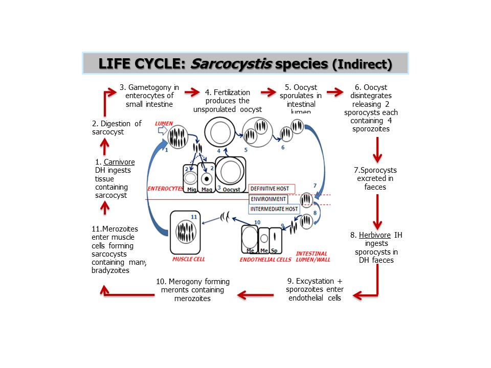
Lifecycle
- Intermediate host (prey) acquires infection by ingestion of sporocyst during grazing or through contaminated feed and water.
- Sporozoites are released after reaching the intestine.
- They penetrate intestinal wall and reach mesenteric lymph node, arteries.
- First generation schizonts develop in endothelial cells of small blood vessels. 2nd generation schizonts develop in vascular endothelium of different organ/tissues including brain and spinal cord.
- Merozoites are released from these schizonts. Merozoites released from 2nd generation initiate sarcocyst formation in straited muscle including cardiac muscles.
- These merozoites first become round to avoid. After repeated asexual division, sarcocyst is filled with crescent shaped bradyzoites which become infective stages on maturation.
- Definitive host acquire infection after ingestion of meal of infected host.
- Bradyzoites are liberated in stomach and intestine. Bradyzoites move actively and penetrate muscosa of small intestine, where they transform to male and female gamonts.
- Macrogamonts are ovoid to round and contain single nucleus. Mature microgamonts are ovoid to elongate and contain microgametes.
- Macrogamete and microgamete fertilize and wall develops around zygote and oocyst is formed.
- Oocyst sporulate in lamina proprid. Sporulated oocyst are then passed out in faceces from which intermediate host gets infection.
- Gametogonic stages develop either in goblet cells at or near the surface of intestine or in enterocysts next to basement membrane adjoining lamina propria or rarely in mesenteric lymph node.
- Because of asynchronous sporulation and cyst wall being membranous, few sporulated oocyst appear in the beginning in faeces followed by discharge of sporocysts for a long period patency.
Mode of transmission
- Transmission in intermediate host is through ingestion of contaminated feed or water.
- Transmission in final host is through ingestion of contaminated meat.
Clinical signs/ Pathogenesis
a. Sarcocystis cruzi (cattle, other species included)
- Fever, Anorexia, Loss of weight, Hair loss, Weakness, Neurological signs and death.
- Muscle twitching, prostration, abortion, reduced milk yield, Hypersalivation.
b. S. tenella (Sheep, other species included)
- Anorexia, Weight loss, fever, Anemia, Loss of wool.
- Abortion, Premature birth, Nervous signs, Myositis, Death.
c. S. capracanis (goat, inckude other species infecting goat)
- Most pathogenic in goats
- Fever, Weakness, Anorexia, Weight loss, Tremors
- Irritability, Abortion and Death.
Sarcocystiosis in Pig
- Three species reported from pigs; miescheriana, S. scichominis, S. porcifelis.
- Clinical signs include weight loss, purpura of skin of ear and buttocks, Dyspnoea, Muscle tremors, Abortion and death.
Sarcocystiosis in Equines
- Four species ; bertami, S. equicanis, S. fayeri and S. asinus
- Clinical signs include anemia, Anorexia, Fever, Excessive salivation, Abortion and Loss of body hair.
Diagnosis
- On the basis of clinical signs.
- Demonstration of sporulated oocyst in feces of definitive host.
- Defection of Ab in serum can help in diagnosis of acute sarcocystiosis. ELISA is used in detection of circulating antigen in mice.
Treatment
- Most of anticoccidials drugs; Amprolium, Salinomycin works.
Prevention and control
- Interruption of life-cycle to minimize spread of sporocyst shed by definitive host. It can be bought by:
- Keeping away carnivorous from animal houses and from their food, water and bedding.
- Uncooked meat should never be fed to carnivorous.
- Dead animals should be buried or incinerated to avoid access to carnivorous.
- Feeding freeze dried meat or cooking meat at 550C for 20 minutes can kill sarcocyst in meat.