Isospora canis
Location and host
- This species is found in small intestine of dog.
Morphology
- Oocyst are ellipsoidal to slightly ovoid, measuring average of 38 x 30 µm.
- Oocyst have smooth pale wall without micropyle, polar granules or residuum but with tiny blob adherent to oocyst wall at broad end.
- Sporocyst are ellipsoidal with smooth colorless wall and a prominent residuum. Size of sporocyst is 18-28 x 15-19 µm.
- Each sporocyst contains four sausage-shaped sporozoites with clear sub-central globules.

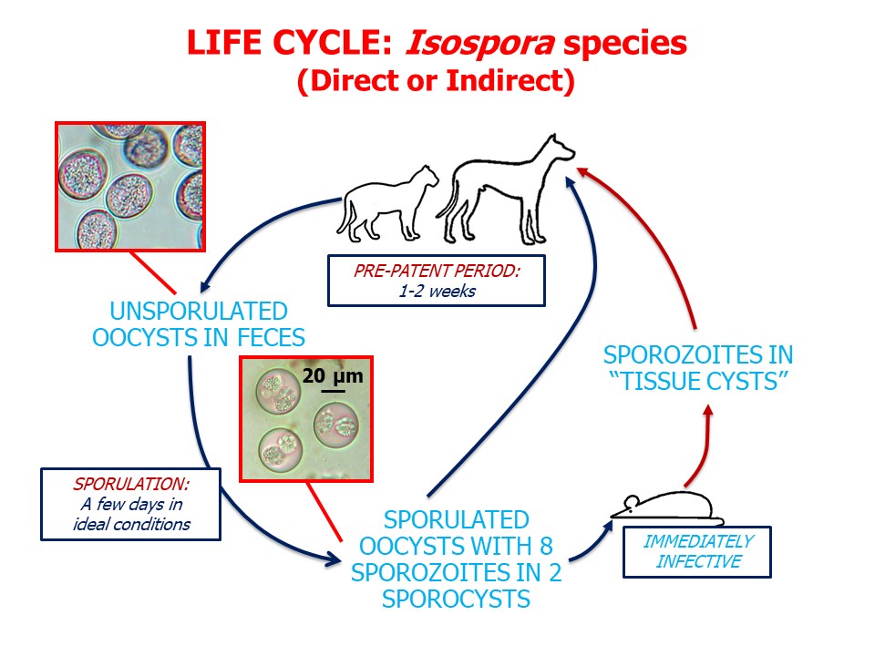
Life-cycle
- Three merogony generations occur in sub epithelium of lamina propria of small intestine.
- Gamonts appears within epithelial cell about7 days post infection.
- Prepatent period : 9-11 days.
Clinical signs/ Pathogenesis
- These species are not pathogenic on their own, but infection may be exacerbated by intercurrent viral disease or other immunosuppressive agents.
- Causes diarrhea in young puppies.
- On PM, villous stunting and reduction in absorptive surface area of lower small intestine may be found.
Diagnosis
- Diagnosis is based on findings of coccidial stages in intestine.
- Finding oocyst in faceces considering presenting signs of sudden onset of enteritis.
Treatment
- Information in treatment is lacking
- Use of sulphonamides such as sukphadimidine should be tried.
Control
- Good sanitation and isolation are effective measure in prevention and control of coccidiosis.
- In kennels or rescue centers, animal accommodation should be cleaned daily.
- Disinfection of kennels, using ammonia based produced are effective in killing oocyst in environment.