Haemoproteus columbae
Location and host: Found in domestic and wild pigeon, doves
Morphology
- Macrogametes and microgametes present in erythrocytes range from tiny ring forms to elongate crescent-shaped that curve around the host cell nucleus in form of halter.
- Macrogametes stain dark blue with Giemsa stain
- Nucleus is red to dark purple and compact and pigment granules are dispersed throughout cytoplasm.
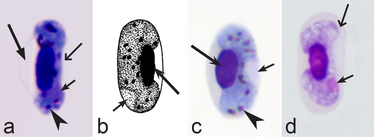
Fig: Haemoproteus columbae elongate gamont surrounding erythrocytic nucleus
Lifecycle
- Birds gets infected when vector flies bites birds for its blood meal. Sporozoites are introduced into host blood.
- Sporozoites after entry into blood stream invades endothelial cells of blood vessel of lungs, liver and spleen.
- Sporozoites undergoes multiple fission under these cells to form schizonts
- Schizonts contain 15 or more unpigmented uninucleate bodies called cytomeres. Each cytomere grows further and undergoes multiple fission. Host cell becomes enlarged and filled with numerous multinuclear cytomeres.
- Each cytomere produce numerous merozoites which break out and pass into blood stream.
- Merozoites enter the red blood cells and form gametocytes. Some merozoites further invade endothelial cells and repeat asexual cycle.
- Gametocytes appear in blood after 28-30 days of infection. Hippoboscid flies when sucks blood from infected host, gametocytes enter flies gut.
- Exflagellation of microgametocyte occurs in gut of flies.
- Fertilization of macrogametocyte with microgametocyte occurs resulting in formation of motile zygote ookinetes.
- Ookinete migrate to outer surface of midgut where sporogony takes place. This results in the production of sporozoites.
- Sporozoites are released into body cavity of flies and passes to salivary gland. These sporozoites then enter host cell through saliva when these flies fly to take blood meal of host.

Mode of transmission : Bite of hippoboscid flies, biting midges (Culicoides), mosquitoes and tabonid flies.
Diagnosis
On the basis of clinical signs
- Affected birds usually show no clinical signs
- In heavy infection, birds appear restless and don’t feed.
- Anemia results due to destruction of erythrocytes.
PM findings
- Liver and spleen may be enlarged
- Schizoints on endothelial cells of lungs, liver, spleen and kidneys may be found
- Demonstration of intraerythrocytic gametocytes in blood smears

Chemoprophylaxis
- Treatment is not known
Prevention and control
- Control of vector flies is sufficient to prevent disease
- Use of fly-repellent may help in prevention of flies towards poultry farm
Other species
a. melagridis
- Macrogametes and microgametes in erythrocyte are elongate and curve around host cell nucleus, occupying half-three quarter of host cell
- Nucleus of microgametocytes is compact
- Denser cytoplasm
b. netlionis
- Synonym H. anatis , H. anseris, H. hermani
- Macrogametes and microgametes are elongated and curve around erythrocyte cell nucleus partially encircling the host cell nucleus and often displacing it.
- Few-30 or more pigment granules which are usually coarse and round and often grouped at the ends of the cell.
- Host cell is not enlarged.
c. sacharovi
- Macro and microgametes completely fill erythrocyte when mature, distorting and pushing the nucleus to one side.
- Pigment granules are sparse compared with other species.