Genus: Leucocytozoan
- These are parasites of birds
- Macrogametes and microgametes reside in leukocytes, some species occasionally in erythrocyte.
- Merogony takes place in parenchyma of liver, heart, kidney and other organs, with meronts forming large bodies (megalomeronts) divided into cytomeres.
- Vectors are black-flies (Simulium) or midges (Culicoides).
Leucocytozoon species
|
Species |
Hosts |
Vectors |
|
Leucocytozoon caulleryi |
Chicken, guinea fowl |
Midges (Culicoides) |
|
L. sabrezesi |
,, |
,, |
|
L. smithi (syn. Leucocytozoon-schueffneri ; L. macleani) |
Turkey |
Black flies (Simulium) |
|
L. Simondi |
Duck, Goose |
Black flies (Simulium) |
|
L. marchouxi (syn. L. tuttur) |
Pigeon, Dove |
Black flies (Simulium) |
|
L. struthionis |
Ostrich |
Black flies (Simulium) |
Morphology
a. caulleryi
- Gamonts are present in erythrocytes. Mature gamonts are round measuring 15.5 x 15 µm and distort the host cell causing host cell nucleus to form a narrow dark band extending about one third around parasite.
- Megalomeronts preset within tissues are 26-30 µm in diameter.
b. sabrezesi
- Gamonts when mature are elongated measuring 22-24 x 4-7 µm.
- Gamonts may distort erythrocyte which becomes spindle shaped with long cytoplasmic horns extending beyond parasite.
- Host cell nucleus form a narrow darkly staining band along one side of parasite.
c. smithi
- Synonyms : L. schueffneri, L. macleani
- Mature gamonts are rounded at first but later become elongate, averaging 20-22 µm in length.
- Host cells are elongate averaging 45 x 14 µm with pale cytoplasmic horns extending out beyond the enclosed parasite.
- Nucleus of host cell are elongated forming a thin dark bond along one side of parasite, often splitting to form a band on each side of parasite.
- Meronts in the hepatocytes of liver are 10-20 x 7-14 µm.
d. simondis
- Mature macrogamonts and microgamonts are elongated, sometimes rounded, 14-22 µm long.
- Gametocytes are present within erythrocytes or leucocytes which become elongate upto 45-55 µm long, with their nucleus forming a long, thin, dark band along one side.
- Infected host cells have pale cytoplasmic horns extending out beyond the parasite and nucleus.
e. marchouxi
- Macrogametes are rounded or elliptical, stain dark blue with Giema stain.
- Macrogametes have complex, reddish nucleus
- These species forms rounded megalomeronts in nearly all internal organs.
f. struthionis
- Gamonts are round and present within erythrocytes.
Lifecycle
a. caulleryi
- Sporozoites are injected into host cell through bite of midges
- Two types of schizonts are developed. First generation schizonts develop in Kupffer cells of liver which are small.
- Merozoites from these small schizont enters either erythrocytes to become gametocytes or enter parenchyma cells of the liver to form hepatic schizonts, having a number of cytomeres.
- After repeated multiple fission, a large number of merozoites develop in lymphoid cells or macrophage in brain, liver, lungs, kidney, intestinal tissue and lymphoid tissues 4-6 days after infection. They contain large number of cytomeres which further produce much larger number of merozoites. These are megaloschizonts.
- Merozoites from hepatic schizonts and megaloschizonts are released into blood and develop as gamonts in peripheral circulation 6-7 days after infection ( inside lymphocytes or in erythrocytes).
- Gamonts are taken up by vector during blood meal. Macrogametes and microgamete fuse to form zygote inside vector.
- Zygote formed is elongated to form ookinete. These passes through midgut wall to form subspherical oocyst on midgut outer wall.
- Sporozoites are formed and passes into salivary glands and introduced into new host when midges bite there.
b. Smithi
- Vector of this parasite is black flies (Simulium sps.)
- Birds acquire infection when infected blackflies bites host.
- Through way of saliva, sporozoites enter blood stream and invade various tissue cells, round up and become meronts.
- Hepatic meronts occur in liver cells containing round crescent shaped basophilic cytomere, which develop into masses of deeply staining merozoites. Merozoites completely fill the host cell cytoplasm.
- Merozoites enters blood cells and forms gamonts.
- Gamonts are taken up by black flies during blood meal. These gamonts invade flies gut, where gametes are formed. Microgametes fuse with macrogametes and form zygote.
- Zygote develops into oocyst to produce sporozoites which breaks oocyst and develop into salivary gland, where they get ready to infect new host, when vectors feed on new host.
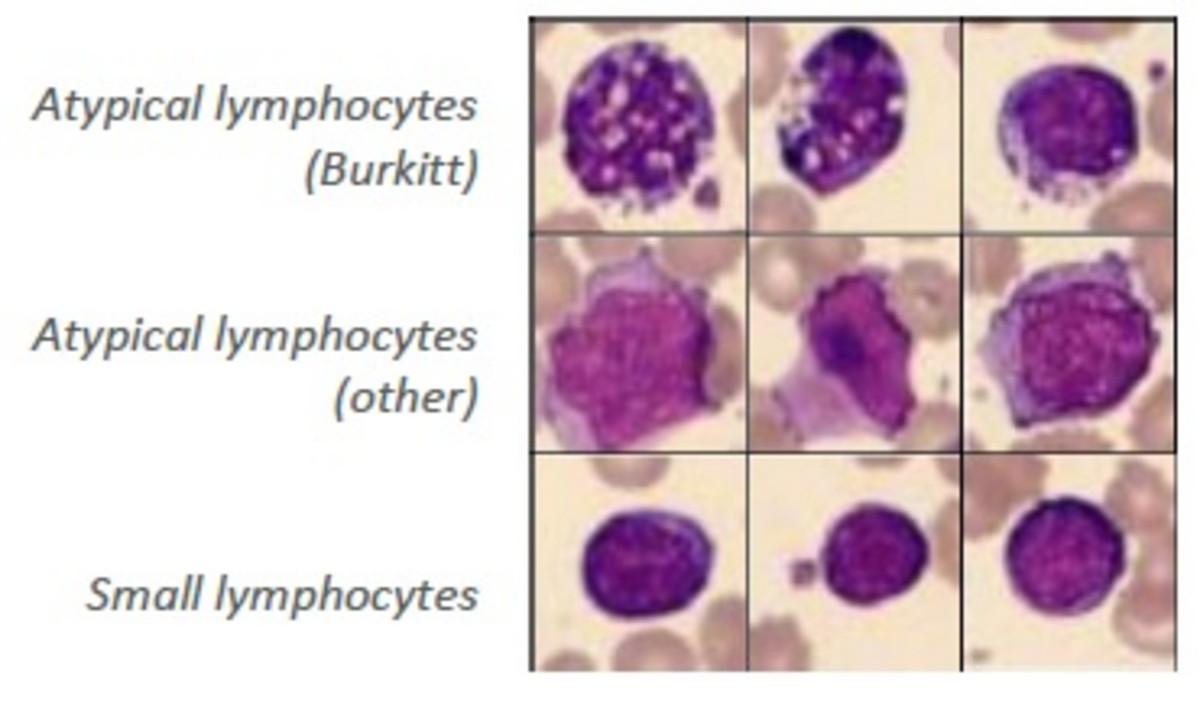
Fig: L. Smithi gamont within an elongated lymphocyte
c. simondi
- Host : Duck, Goose
- Vector: Black flies (Simulium).
- Birds acquire infection when black flies bite them.
- Through way of saliva, sporozoites enters blood stream of host.
- They invade various tissue cells, round up and become meronts.
- Two types of meronts occur; hepatic meronts, occurring in liver cells and megalomeronts found in brain, lungs, liver, heart, kidney, gizzard, intestine and lymphoid tissues 4-5 days after exposure.
- Hepatic meronts forms a large number of cytomeres which undergoes multiple fission to form small merozoites. On other hand, megalomeronts produce thousands of bipolar merozoites.
- Merozoites enters blood cells and form gamonts. Merogony continues in organ for indefinite time, but at reduced rate during this phase, birds are source of infection for new crop of ducklings.
- Gamonts are then taken up by black flies when bites host.
- Macrogametes and microgametes are formed inside gut of vector. 4-8 microgametes are formed by ex-flagellation from microgamonts.
- Microgamete and macrogamete fuse to form motile zygote or ookinete. Ookinete develop into oocyst inside gut wall. Oocyst forms several sporozoites (5-10 µm long, one end rounded and other pointed).
- Sporozoites breaks out oocyst and passes into salivary glands of insects from where they infect new host, when vector visits host to take blood meal.
Pathogenesis/ Clinical signs
- Young birds suffer more than adults.
- Clinical signs include: Anaemia, List lessness, Loss of appetite and greenish diarrhoea
- PM findings include : Haemorrhages in lungs, liver and kidneys, Enlargement of spleen and white spots on heart muscles.
Diagnosis
- On the basis of clinical signs
- On the basis of PM findings
- Demonstration of gamonts in chicken erythrocyte.
Chemoprophylaxis/ Treatment
- Sulpha monomethoxine sodium @ 1 g/liter of drinking water
- For prophylaxis, continuous medication @ 1g/20 liters of drinking water.
Prevention and control
- Control of vector flies using flies repellant may be effective in prevention of disease.