E. zuernii
Location and host: Found in small and large intestine of cattle.
Identification features/ Morphology
- Oocyst are sub-spherical, colorless, measuring average size of 17.8 x 15.6 µm, with no micropyle or oocyst residuum.
- Sporocyst are ovoid, 7-14 x 4-8 µm, each with tiny stieda body and sporocyst residuum is usually absent.
- Sporozoites are elongate and lie head to tail in sporocysts, each has a clear globule at the large end.

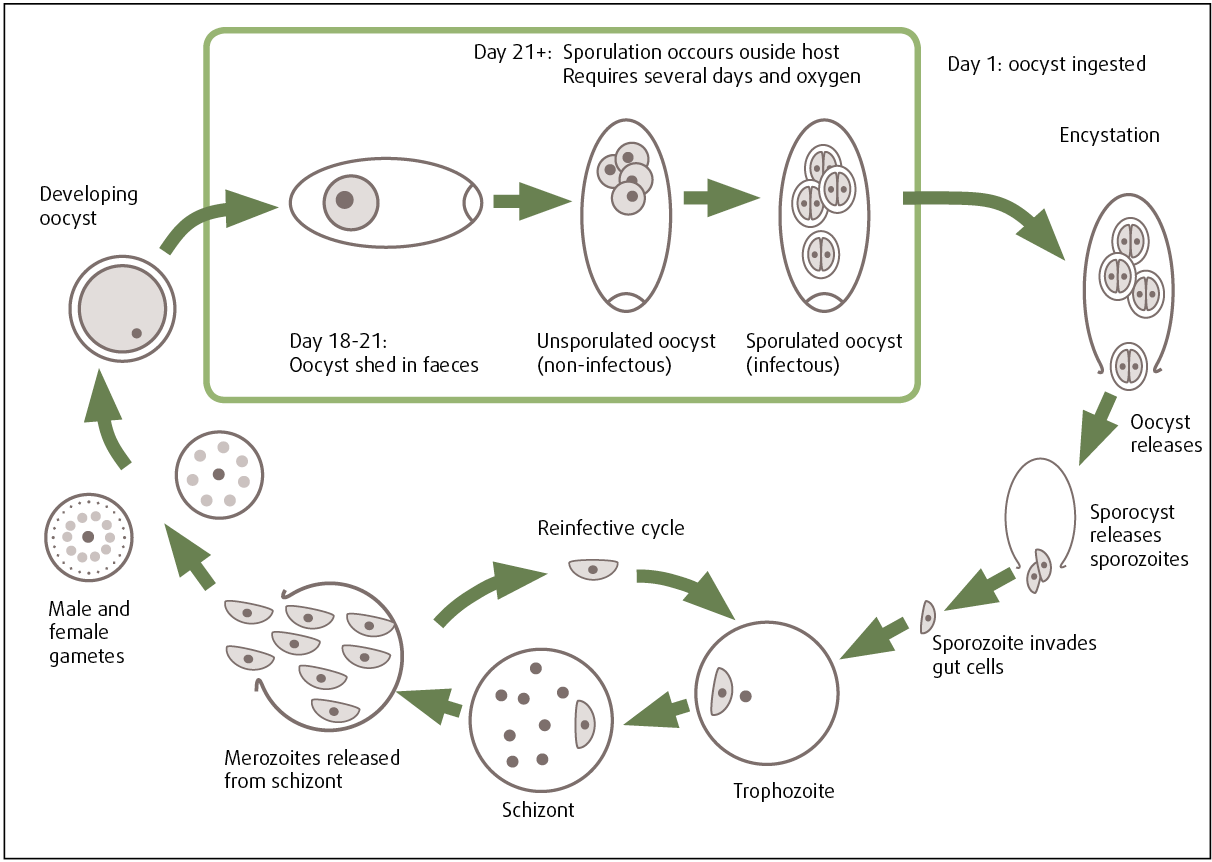
Life cycle
- Two asexual generation occurs; first generation meronts are found in lamina propria of lower ileum and mature at 14-16 days after infection.
- Second generation meronts occur in epithelial cells of caecum and proximal colon.
- Sexual stages occurs generally in epithelial cells of caecum and colon.
- Prepatent period: 15-17 days
- Patent period: 5-17 days
- Sporulation time: 2-10 days
Clinical signs / Pathogenesis
- Acute infection of zuernii, causes hemorrhagic diarrhea in calves. First, feces are streaked with blood, but as diarrhea become more severe, bloody fluid, clots of blood and liquid faeces are passed.
- Tenesmus and coughing can result in diarrhea being spurted out upto 2-3 m.
- Hind quarter of animals are smeared with diarrhea.
- Affected animals shows emaciation, dehydration, weakness, listlessness. Rough coat, sunken eyes and drooping ear may be seen.
PM findings
- Generalized catarrhal enteritis.
- Lower small intestine, caecum and colon may be filled with semi-fluid hemorrhagic material.
- Large or small areas of intestinal mucosa may be eroded and destroyed.
- Mucous membrane may be thickened with irregular whitish ridges in large intestine or smooth dull-grey areas in small intestine or caecum.
- Diffuse hemorrhage in intestine in acute cases and potential hemorrhages are seen in milder cases.
Diagnosis
- Diagnosis is made on basis of clinical signs; bloody diarrhea, emaciation, dehydration, tenesmus.
- Demonstration of oocyst in faeces.
- On basis of histopathological findings; small intestine, caecum may be filled with semi-fluid hemorrhagic material.
Treatment
- Treatment is done through use of sulphonamide drug
- Amprotium-20-30 mg/kg body weight, daily in feed for 4-5 days.
- Lincomycin HCl – 1gm/ calf given in drinking water for 21 days.
Prevention and control
- Same as in bovis