E. tenella
- Most common and highly pathogenic coccidium of domestic fowl.
- Primary agent for caecal
Location
- Occurs in small intestine of poultry
Morphology
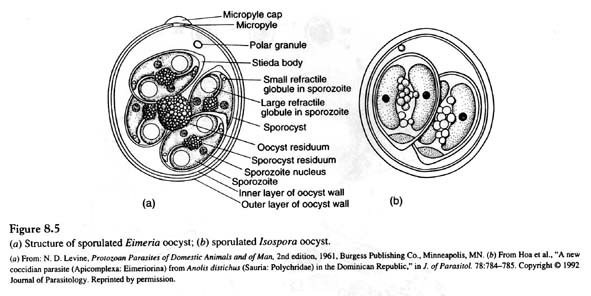
- Oocyst are ovoid, smooth, colorless without micropyle or residuum but with polar granule
- Average size of oocyst 25 x 19 µm.
- Sporocyst are ovoid
- Stieda body is indistinct
Life cycle
- All endogenous stages occur in endothelial cells of villi and crypts of caeca.
- Two schizogonic generations of large epithelial schizonts developing in cells of crypts and third generation of small epithelial schizonts develop along with gamonts in the cells of whole mucosa of caeca.
- Prepatent period is 6 days.

Clinical signs
- tenelia is most pathogenic coccidium and responsible for caecal coccidiosis.
- Clinical signs appear after 4th day of infection.
- Clinical signs exhibited by infected includes: Poor growth, emaciation, poor egg production, loss of appetite, Restlessness and drooping wings, sluggish movement in 6th day, Sulphur diarrhea or large quantities of blood seen in case of diarrhea.
PM findings
- Petechial hemorrhage is seen on caecum during first three day which become hemorrhagic by fourth day.
- Caeca may be swollen or enlarged; wall may be thickened.
- Caeca are filled with large amount of unclotted blood or partly clotted blood on 5th day of infection.
- On 7th day of infection, caeca is filled with fibrinous and necrotic materials and large number of oocysts.
Diagnosis
- History of flock, characters of various types of lesions and their location, important features of life-cycle are essential.
- Diagnosis is usually made on basis of clinical signs, PM findings and demonstration of oocyst in feces.