E. necatrix
Location and host
- This parasite is found in chicken throughout world. It occurs in jejunum, mid-gut, caecum or other parts of large intestine.
Morphological features
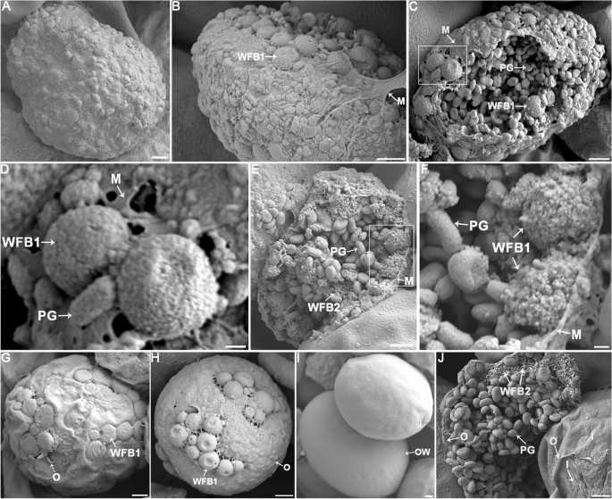
- Oocyst are ovoid, smooth, colorless, measuring an average of 20 x 17 µm.
- Micropyle or residuum are absent but polar granules are present.
- Sporocyst are ovoid, with a stieda body and without a residuum.
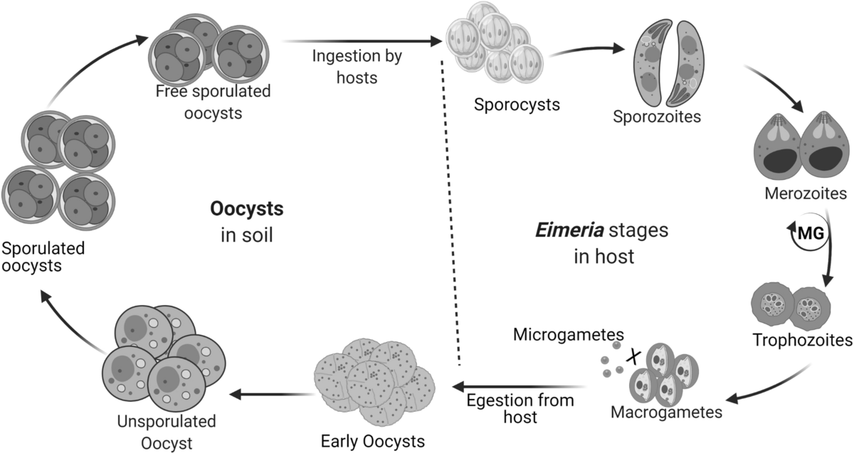
Life cycle
- After ingestion of sporulated oocyst and excystation, sporozoites enter epithelial cells of small intestine, pass through epithelium into lamina propria at center of villi and migrate towards muscular mucosae.
- Many sporozoites are engulfed by macrophages during this passage and are transported to epithelial cells of the fundus.
- Sporozoites round up to form first-generation meronts above host cell nuclei in epithelial cells of crypts of intestine.
- Second generation meronts develop deep in the mucosa.
- Third generation is small epithelial schizont which develops in large number along with gamonts.
- Pre patent period: 138 hours (6-7 days)
- Patent period: 12 days
- Sporulation time: 18-24 hours.
Clinical signs/ pathogenesis
- Most pathogenic species of poultry causing chronic intestinal coccidiosis. Mostly middle 3rd of small intestine is affected.
- Clinical signs exhibited by affecting birds include:
- Diarrhea ( mucoid and sometimes bloody)
- Dejection ( sad and depressed state)
- Ruffled feathers
- Drooping wings
- Inappetance
- Weight loss
- Depressed weight gain
- Death occurs after 5-7 days of infection, before oocyst pass in faeces.
- Recovered birds remain unthrifty and emaciated.
PM findings
- Intestinal lesions are usually found on middle third.
- Small white opaque foci may be seen after fourth day of infection. These are 2nd generation meronts.
- Severe hemorrhages occur by day 5 or 6 and small intestine may be markedly swollen and filled with clotted and unclotted blood.
- Intestinal wall is thickened and dull red and petechiae are present in white foci.
- Gut wall may lose its contractility, become friable and epithelium may slough and replaced by network of fibrin containing mononuclear cells. This network is replaced by connective tissue resulting in permanent scarring, which interferes intestinal absorption.
- Lesion are sored +1 to +4 as follows:
- +1 presence of small scattered petechiae and white spots visible from serosal surface.
- +2 numerous petechiae on serosal surface and some slight ballooning of intestine.
- +3 extensive hemorrhage in lumen and presence of red or brown mucus, extensive petechiae on serosal surface, marked ballooning of intestine and absence of normal intestinal contents.
- Ballooning may be extensive and hemorrhage may give an intensive dark color to intestinal contents.
Diagnosis
- Diagnosis of coccidiosis is usually based on clinical signs, PM findings and demonstration of oocyst in faeces of infected birds.
Treatment ( All coccidia)
- Sulphanamide drug have been mostly used and recommended that they can be given for two periods of 3 days in drinking water with interval of 2 days between treatment.
- Amprolium mixed with sulphaquinoxaline at levels of 0.006% of each in food is more effective against poultry coccidia.
- Amprolium powder or amprosol 1 gm dissolved in 1 liter of water for 5-7 days.
- Coxiquin or Duoxoxin 1 gm powder dissolved in two liters of water for 5 days.
- Codrinal powder 4 gm dissolved in 1 liter of water for 3-6 days
- Coximar 30 gm/ 30 liter water for 3 days
- Vitamin A supplements for 3-5 days in water. Kaysol Forte 5 gm for 200 birds, liquid 5ml/100 birds or 10 ml/100 birds.
Prevention and control measures of poultry coccidiosis
- Destruction of oocyst: it can be achieved by following methods:
- Fumigation of poultry houses and litters by 0.044% solution of ammonia. It will kill oocyst within 2 hours.
- By application of methyl bromide @ 1 lbs/1000 sq. ft of poultry litter.
- Proper sanitation and management should be adopted in poultry houses.
- Young birds should be kept separately from adult ones.
- Poultry litters should be kept dry and stirred frequently.
- In case of outbreak, sick birds should be removed immediately and kept in separate house. Remaining healthy birds should be treated with coccidiostats and infected litter should be changed.
- As oocyst can be carried by cloths of attendants of poultry house, shoes should be removed outside of poultry house before entering into it.