E. histolytica
Location and host
- Found in caecum and colon of man, pig, dog, cat. Monkey and sometimes rat.
- This protozoan caused disease called amoebic dysentery or amoebiasis.
Morphology
- Parasite has two forms: Trophozoite form and cyst form.

Trophozoite form
- These occur in two varieties: large form and small form. Large forms are more pathogenic and size is more than 20-30 µm in diameter.
- Movement of trophozoite is through pseudopodia and movement is rapid in warm environment. Movement is crawling type.
- Cytoplasm has clear ectoplasm or endoplasm. Endoplasm has food vacuole which contain host cell, RBC and sometime bacteria.
- Nucleus is spherical and measure 4-7 µm in diameter. They are indistinct in living amoeba. Nucleus has small central endosome with ring of small peripheral granules. Nucleus membrane is lined by a row of fine chromatin granules.
Cyst form
- Encystation of trophozoite starts with dehydration of faeces in lumen of colon. Trophozoite discharge undigested food and condenses into a spherical mass called cyst.
- Cyst matures by mitotic division of nucleus and form four nuclei. Rarely eight nuclei may be found.
- Cyst are spherical or sub-spherical in shape and size is about 5-20 µm in diameter.
- Cyst have double-layered cyst wall and passed out in constipated faeces.
- Immature stages contain 1-2 nuclei, glycogen granules and chromatoid rod acts as reserve food material. Chromatid rods are absent in mature stages.
- Cyst can survive for several days at 4-8 0 These are sensitive to putrefication, desiccation and temperature above 400C or below -50C. Chlorination of water does not kill the cysts, buy 5% acetic acid kills them in a few minutes.
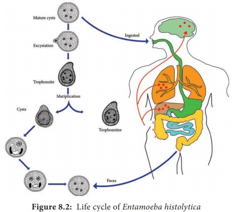
Life cycle
- Host gets infection when they ingested mature cyst with contaminated food and water.
- After ingestion, cyst reaches intestine of host, where they multiply and divide. Four nuclei forms at first and later eight nucleated forms are formed.
- Cyst then ruptures and these nucleated bodies come out which are known as amoebulae. They further pass into large intestine and develop into trophozoites.
- Trophozoite multiplies by binary fission and penetrate the wall of large intestine by means of pseudopodia.
- Cyst formation then takes place before encysting amoeba rounds up, eliminate its food vacuoles and becomes smaller.
- Cyst wall develops and nucleus divides into two and then into four nuclei. Cyst are passed out in faceces and hosts gets infection when they ingest these cysts.
Mode of transmission
- Through ingestion of contaminated food and water that contains uninucleate cyst.
- Lack of personal hygiene, use of water polluted by night soil.
Pathogenesis / Clinical signs
- Amoebiasis in man is common and serious disease caused by large form of histolytica. E. coli or Aerobacter aerogens play important role in growth and invasive character of amoeba.
- Disease may be acute or sub-acute, but chronic infections are most common.
- Parasite penetrate intestinal wall by lysis of epithelium.
- Amoeba multiplies in epithelium forming small colonies and then penetrate sub-mucosa.
- They invade deep into mucus membrane and produce inverted flask-shaped ulcer, having narrow canal or neck opening into bowel lumen. Amoeba are mainly found at periphery of ulcer and cavity of ulcer is filled with necrotic tissues.
- Amoeba may reach other parts of body through lymph or blood vessels. In liver, they produce inflammation and abscesses. They may be carried to lungs and brain by blood.
Clinical signs
- Clinical signs may be intestinal form and extra-intestinal form.
Intestinal forms
- It shows acute and chronic forms.
In acute form
- there is dysentery. Faeces consist of almost entirely of blood and mucus with amoeba.
- Severe abdominal pain with straining.
- Loss of appetite
- Nausea and vomiting
- Fever may be seen.
In chronic form
- Recurring symptoms occur for long time
- Occasional abdominal pair
- Nausea and vomiting
- Flatulence
- Irregular bowel
- Headache and fatigue
- Symptoms stimulate appendicitis and peptic ulceration. Rectal straining may cause prolapse of rectum.
- Pigs also show similar symptoms
- Dogs may have lesions in caecum and mostly without symptoms, but they may suffer from acute or chronic amoebiasis
- Kitten shows acute dysentery with extensive intestinal ulceration.
Extra -intestinal form
- Abscess in liver, may produce pain in liver.
- High fever
- Increase in leucocyte count
- Brain and lung abscess in some cases
Diagnosis
- On the basis of clinical signs and lesions
- Detection of cyst or trophozoites in faceces.
- Faecal culture to demonstrate amoeba
- Liver abscess detection with help of x-ray
- Blood sample count shows increase in leucocyte count
- Serological test : CFT.
Treatment
- Metronidazole is drug of choice. It is given @ 750 mg three times a day for 5-10 days. Tinidazole @ 2 gm once a day for 3 days can be given as alternative to metronidazole.
- Emetine hydrochloride 65 mg dissolved in 1 ml of distilled water and provide deep intramuscular or subcutaneous injection for 5-10 days in human and dog for extra intestinal form of disease.
- Chloroquine 300 mg once daily, orally for 5 days.
Control measures
- Adoption of proper sanitation, personal hygiene
- Use of clean drinking water
- Proper disposal of sewage and night soil
- Prevention of fecal contamination of food by flies.