E. bovis
Location
- They are usually found in small and large intestine of cattle.
Identification/ Morphology
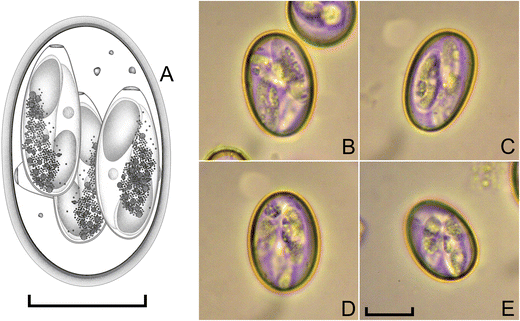
- Oocyst are ovoid or subspherical, colorless measuring average of 27.7 x 20.3 µm.
- Oocyst have smooth wall with inconspicuous micropyle, no polar granule or oocyst residuum.
- Sporocyst are elongate, ovoid, 13-18 x 5-8 µm and have inconspicuous stieda body and sporocyst residuum.
- Sporozoites are elongate and lie length wise head to tail in the sporocyst and usually have clear globule at each end.
Lifecycle
- Two asexual generation , first generation occurs in endothelial cells of the lacteals of villi in posterior half of SI and mature at 14-18 days of infection.
- Second generation occurs in epithelial cells of cecum and cecum or colon but may extend into meter of SI in heavy infection.
- Sexual stages generally occur in caecum and colon.
- Prepatent period: 16-21 days
- Patent period: 5-15 days
- Sporulation time: 2-3 days.

Ingestion of sporulated oocyst by host —- > Release of sporozoites in endothelial cells of villi. First generation schizogony —- > First generation schizont invades another cells, epithelial cells of villi — > Release of merozoites — > Merozoites undergoes gametogony —- > Formation of zygote from fertilization of male and female gamete — > Oocyst forms and released in feces — > Sporulation in environment in optimum condition —- > Repeat
Clinical signs and pathogenesis
- Severe diarrhea usually bloody diarrhea (foul-smelling). Diarrhea may also contain masses of mucus and clots of blood.
- Enteritis
- Tenesmus in case of heavy infection.
- Animal may be pyrexic, weak and dehydrate if untreated, they may die.
- Rough coat, dropping ears, listlessness, soiled hindquarters, anorexia, inability to rise ad partial paralysis of anal sphincter in case of mixed infection with zuernii.
PM findings
- Mucosa of ileum appears congested, edematous and thickened with petechiae or diffuse hemorrhage.
- Gut lumen may contain large amount of blood.
- Sloughing of mucosa in chronic case.
Diagnosis
- Diagnosis is usually made through its characteristics symptoms, foul-smelling diarrhea, tenesmus, loss of appetite.
- Demonstration of oocyst in faeces.
- PM findings: characteristic intestinal lesions.
Treatment
- Treatment is usually through sulphonamide, such as sulphodimidine or sulphamethoxypyriazne given orally or parenterally. This is repeated at half the initial dose level on each of next 2 days.
- Though clinical signs are seen only when disease is advanced, these drugs are not effective as much.
Control
- Prevention and control is based on good hygiene or management.
- Feed trough and water containers should be cleaned regularly.
- Bedding should be kept dry.
- Frequent fecal examination should be carried out.
- Calves should be kept isolated within 24 hours after birth. Growing calves should not be kept with adult animals.