Babesia
- It is worldwide in distribution and has 70 species recorded from different categories of domestic animals.
- Causes tick-borne haemoprotozoon disease “Babesiosis” in cattle, sheep, goat, horses, pigs and dogs.
Morphology
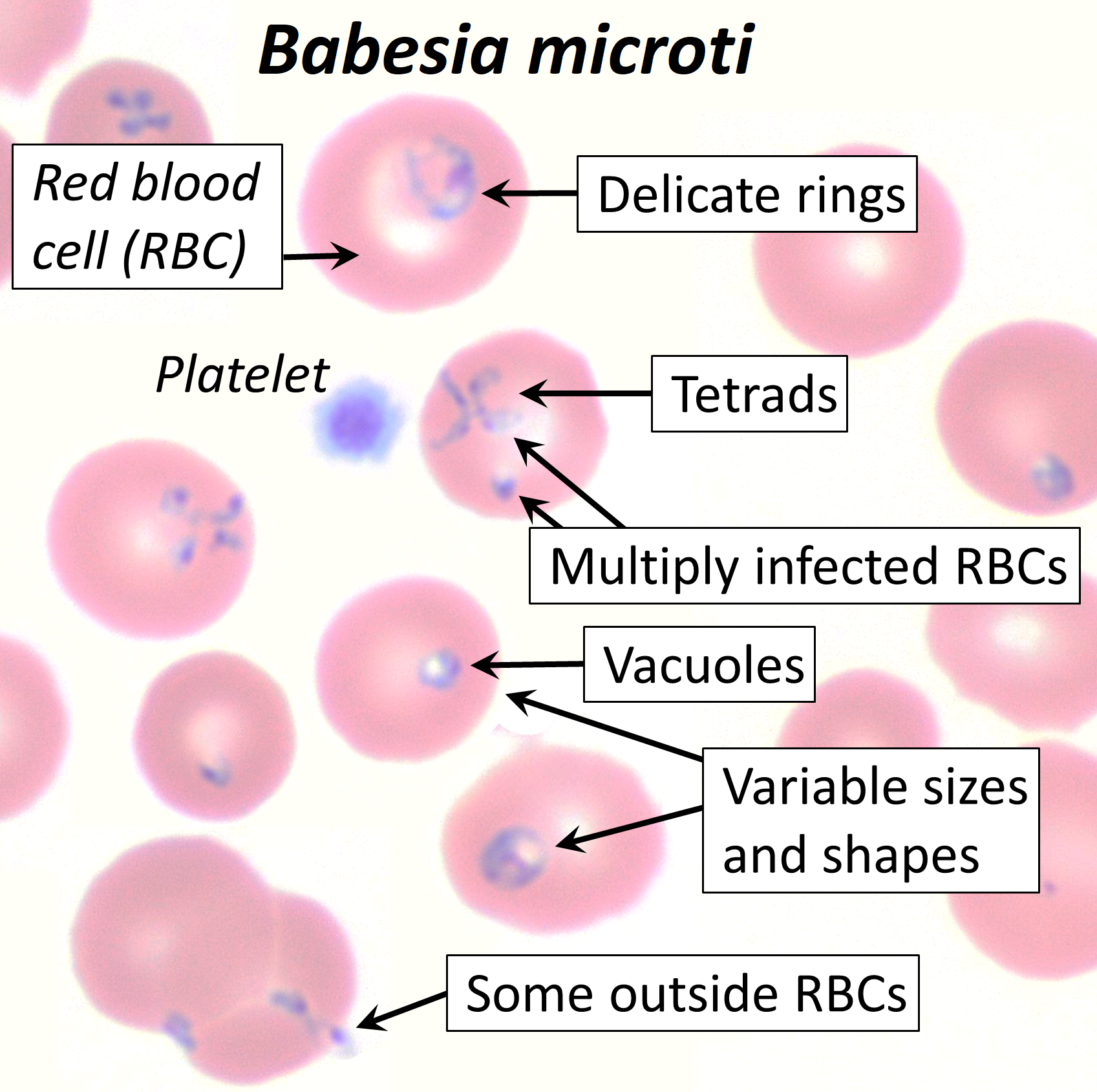
- Organisms multiply in erythrocytes by asexual division, producing two, four or more parasites without pigments.
- They are found singly in RBC as round, ovid, elongate or amoeboid trophozoites, in pairs as pyriform merozoites or in tetrads as cruciform merozoites.
- Characteristically, they are pear-shaped lying at an angle with narrow ends in opposition ( side by side)
- Generally, Babesia are divided into two major groups:
|
Large form |
Small form |
|
Average length of more than 3 µm. |
Average length of less than 2.5 µm. |
|
In paired forms and lie with their narrow ends at an acute angle. |
Lie at an obtuse angle. |
|
Infection with large form can be successfully treated with trypan blue. |
Infection with these forms don’t respond to trypan blue. |
Life cycle
- These parasites have heteroxenous cycle: asexual stage occurs in vertebrate host and sexual stages occur in invertebrate host ( ticks).
- Babesia occur in erythrocytes of vertebrate host when infected ticks feed on them.
- In vertebrate, they multiply by binary fission or budding process or to form two, four or more trophozoites.
- Trophozoites gets released from infected cells and invade other cells. This process repeated until large number of RBC are parasitized.
- Ticks now gets infection when they feed on infected host. Development and transmission in ticks is either ‘transovarian’ or by ‘stage to stage’.

Transovarian transmission
- Occurs only in one-host ticks. Example Boophilus.
- After repeated multiplication of parasite inside erythrocyte of vertebrate, stage are differentiated as micro or macrogametes.
- These stages are taken up by tick when they feed on host.
- Babesia reaches gut of ticks ad starts further development. Gametes are released through lysis of infected erythrocyte and unite to form zygote.
- Zygote undergoes further development and multiplication. They become motile and comes to serous surface and ruptures.
- Vermicules isogametes are released from zygote. These vermicules invade ova of ticks and reach to gut epithelium of larva emerging out of infected eggs.
- Infective organism then reaches to salivary gland of larva and after maturation, they are able to infect vertebrate host during feeding on its blood.
Stage to stage transmission
- Multiplication of developmental forms was seen in phagocytes.
- ‘Pseudocyst’ organism occurs about 7 days after nymph drops off the infected host.
- In those cycle, club-shaped organism of 9×2 µm are formed by 11-15 days.
- These are then liberated from host cell and migrate to the muscle sheath of nymphal tick where they penetrate muscle cells and divide repeatedly to form large number of small ovoid forms.
- After molting of nymph to adult, these organisms migrate to salivary gland of tick feed on host.
- On reaching salivary glands, parasite enter the cells of acini and undergo repeated binary fission to form large number of small, ovoid infective stages.
Babesiosis in cattle and water buffalo
- Four species of Babesia occur in cattle
- Babesia bigemina
- bovis
- divergens
- major