Transmissible Venereal Tumors
Also known as infectious or venereal granuloma, Sticker tumor, transmissible sarcoma, and contagious venereal tumor

Etiology :
- The transmissible venereal tumor is transmitted to the genitals by coitus
- The origin of this tumor is still unknown but has been described as a tumor of lymphocytes, histiocytes, and reticuloendothelial cells.
- It is most prevalent in temperate climates
Clinical signs.
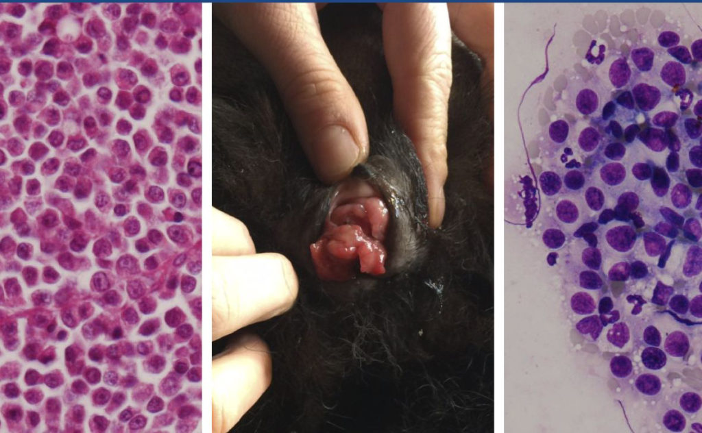
- The tumors are usually cauliflower-like masses on the external genitalia, but they can also be pedunculated, nodular, papillary, or multilobulated.
- These friable masses vary in size up to 10 cm, and hemorrhage is frequently observed. In male dogs, the lesions are found on the caudal part of the penis from the crura to the bulbus glandis or on the glans penis
- Less frequently, the tumor is found on the prepuce.
- Females typically have lesions in the posterior vagina at the junction of the vestibule and vagina.
- When located around the urethral orifice, the mass may protrude from the vulva.
- These tumors have also been reported in the oral cavity, skin, and eyes.
Epizootiology and transmission.
- Transmissible venereal tumors are most commonly observed in young, sexually active dogs.
- Transmission takes place during coitus when injury to the genitalia allows for transplantation of the tumor.
- Genital to oral to genital transmission has also been documented
- Extragenital lesions are believed to be a result of trauma prior to exposure to the tumor.
- Al;so transferred by licking , sniffing or parturition
Pathological finding :
- Histologically, cells are arranged in compact masses or sheets.
- The cells are round, ovoid, or polyhedral and have large, round hyperchromatic nuclei.
- Also present are large nucleoli and numerous mitotic figures.
- The cytoplasm is eosinophilic, and cell borders are indistinct.

Pathogenesis.
- Tumor growth is rapid after implantation but later slows.
- Metastasis is rare (<5% of cases) but may involve the superficial inguinal and external iliac lymph nodes as well as distant sites.
How does CTVT escape the immune system ?
- CTVT is a foreign graft within the host
- The mechanism used by CTVT to escape the immune system in its allogeneic host are incompletely understood; however downregulation of major histocompatibility complex (MHC) molecules and recruitment of an immunosuppressive microenvironment probably plays a role .
Diagnosis :
- Cytology / histopathological examination of FNA
- Signs and lesion
- PCR
DDX :
- Lymphomas
- Histiocytomas
- mast cell tumors
- amelanotic melanomas