Testicular tumors :
A testicular tumor is a tumor that develops from a disordered
uncontrolled growth of cells within the testicles.
- The three most common types of testicular tumors develop from germ cells (cells that make sperm), Leydig cells (cells that produce testosterone), and Sertoli cells (cells that help in the development of sperm):
- Seminomas develop from germ cells
- Interstitial cell tumors develop from Leydig cells
- Sertoli cell tumors develop from Sertoli cells

- Other types of tumors may develop from other cells within the testicles, but these types are rare. Testicular tumors are considered one of the most common tumors in older intact (not neutered) male dogs and are rare in cats.
Etiology :
- The reason is not straightforward.
- Very few tumors and cancers have a single known cause.
- Most seem to be caused by a complex mix of risk factors, some environmental and some genetic or hereditary.
- In testicular tumors, cryptorchid pets have a tendency to develop Sertoli cell tumors and seminomas, but not interstitial cell tumors.
- Older cryptorchid dogs (greater than 6 years of age) have a much higher tendency to develop some form of testicular cancer; older dogs in general have a higher rate of developing these tumors.
Progression of tumors :
- These tumors typically are locally a problem, meaning they have a low rate of spread.
- Both Sertoli cell tumors and seminomas have less than a 15% chance of spread to lymph nodes or other organs.
- Interstitial cell tumors rarely spread.
- Because other cancers of the urinary or reproductive system may spread to the testicles, staging is imperative.
Clinical signs :
- Mostly it do not have any obvious clinical signs but vary depending on the type of tumor and the tumor location
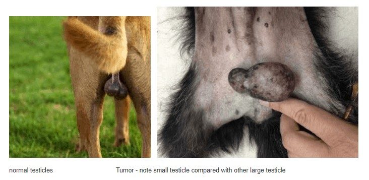
- The clinical signs may be limited to the presence of a mass (or masses) within the affected testicle.
- Palpation of the scrotum may reveal a nodular enlargement of the testicle, unevenly sized testicles, or generalized swelling of the scrotum.
- Sertoli cell tumors can produce estrogen and a condition called hyperestrogenism. Hyperestrogenism can cause signs of feminization.
- This includes :
- enlarged mammary glands and nipples
- a pendulous prepuce
- hair loss and hyperpigmentation (darkening) of the skin.
- Excess estrogen can also depress the bone marrow, causing anemia (pale gums) and lethargy.
- In the rare case of a malignant tumor, the signs may be related to the organs to which the tumor has spread, and can include weight loss, decreased appetite, lethargy, or vomiting.
- If the tumor has metastasized to the lymph nodes near the urinary system or the prostate gland, signs may include difficulty urinating or defecating.
Diagnosis :
- Routine physical examination
- Histopathology
- Cytology
- Fine needle aspiration (FNA)