Pullorum disease
- The historical name is bacillary white diarrhoea.
- Pullorum disease is characterized by very high fever in young chicken and turkeys. Affected birds huddle near the heat source, they are anorectic, weak , depressed , and have white fecal material pasted in the vent area.
- Birds may have respiratory disease, blindness or swollen joints.
Etiology:
Salmonella enterica Pullorum usually causes mortality in young chicken and turkeys within the first 2-3 weeks of age and also affects other chickens and domestic fowl.
- Salmonella : Rod-shaped, Gram-negative, Facultative anaerobic, Non-spore-forming
It belongs to the same family as Escherichia, which includes the species E.coli.
Transmission :
- Vertical ( transovarial) but also occurs via direct or indirect contact with infected birds ( respiratory or fecal)
- Through contaminated fed , water , or litter
- Egg or hatchery infection
- Transmission between farm is due to poor biosecurity
Pathogenesis :
Infection with high or infective dose of S. pullorum via oral route
⬇
Bacteria invades digestive epithelia ; irritation and causes diarrhoea
⬇
Ultimately enters into blood causing bacteremia.
⬇
Bacteria seeded into cells and tissues of different organs (liver, lung, spleen, kidney , different parts of the reproductive tract of hens and testes of male ) causing pathological lesions.
⬇
The bacteria invade ovary and egg follicle and transmit into laid eggs then to hatched chicks.
Lesions:
- Gross lesion :
- No lesions occur in acute cases due to septicaemia and death.
- Gross lesions in adults are not generally seen.
- Lesions in young include :
- Unabsorbed yolk sac.
- Classic grey nodules in liver , spleen , lungs, heart , gizzard , intestine , etc .
- Synovitis
- Gross lesions in adults are not generally seen .
- Deformed ova with atrophic follicles.
- Pericarditis
- Articular and peri articular swelling of hock and wing joint ( arthritis)
- Tumor like lesion in gizzard and heart
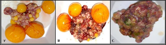
Fig: misshapen ovaries with atrophic follicles.
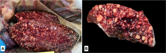
Fig : granulomatous hepatitis( small necrotic foci )

Fig: granulomatous lesion in spleen(raised white spot)

Fig : granulomatous lesion in ovary ( oophoritis)
2. Microscopic lesion:
- firm, cheesy material in the caeca ( caecal core ) and raised plague in the mucosa of lower intestine .
- nodular pericarditis
- Fibrinous peritonitis or haemorrhagic
- Regressing ovarian follicle with caesous content.
- Chronic infection produces lesions indistinguishable from those of fowl typhoid.
Clinical sign :
- Droopiness
- ruffled feather
- chilled appearance with birds huddling near source of heat
- labored breathing or respiration distress
- presence of white diarrhoea pasted around the vent known as “pasted vent”.
- Pot bellied
- Loss of appetite
- Depression
- Pale shrunken comb
- Low egg production
Diagnosis :
- History
- Clinical signs and lesions
- Postmortem finding
- Isolation and identification of bacteria