Necrotic enteritis :
- An acute bacterial infection primarily of chickens and turkeys,although other avian species can be affected.
- Characterised by sudden death, friable and distended intestines and severe necrosis of intestinal mucosa.
- The disease primarily affects broiler chicken ( 2-5 w) and turkey(7-12 w old) .
- Infection occurs by faecal-oral route.

Etiology :
Clostridium perfringens Type ( A or C) and their toxins ( alpha- toxin and sialidase )
- Gram positive
- Obligatory anareobe
- Non- motile
- Rod shaped
- Spore forming bacterium ( spores are highly resistant )
- perfringens are ubiquitous and are a normal inhabitant of the intestinal tract .
Transmission :
- Through contaminated food and water
Predisposing factors :
- Small intestinal coccidiosis
- Diet high in cereal grains
- High amount of animal protein
- Animal fat
- Infection of chickens with immunosuppressive viruses.
Pathogenesis :
Organism enter into body via oral route
⬇
It reaches to the intestine
⬇
Proliferates in intestinal tract
⬇
Produces potent toxins that severely damage the intestinal mucosa
⬇
Toxins absorbed from intestinal tract resulting in toxaemia , which is responsible
for death of body
Thus, NE is a type of enterotoxemia.
- perfringens divided into 5 toxinotypes ( A,B,C,D,E) based on four major toxins – alpha, beta, epsilon and iota .
- The majority of isolates from NE cases are type A ; few caused by type C.
Alpha – toxin produced by Type A&C and beta- toxin by Type C
Toxin / virulence factor :
- 𝛼- toxin produced by TypeA as well as four other toxinotypes are important virulence factors in the pathogenicity of organism .
- 𝛼- toxin is a zinc metalloenzyme with phospholipase activities.
- It hydrolyses phospholipids in membranes of RBC & WBC , thrombocytes, endothelial cells, and muscle cells.
- Thus, toxin is hemolytic, cytotoxic, necrotizing and potentially lethal.
Clinical signs :
- NE has a short clinical course :
Birds are found dead without premonitory clinical signs.Birds appear listless and lethargic for a
a few hours before death.
Mortality in broiler is usually below 10% , but can be as high as 50%.
- Mild and subclinical NE :
Birds may not die but show reduced weight gains,higher FCR.
- Typical signs :
- Depression
- Reluctance to move
- Watery diarrhoea
- Ruffled feathers
- Somnolence
- Decreased appetite / anorexia
- Dehydration
- Huddling
- Drooling of saliva

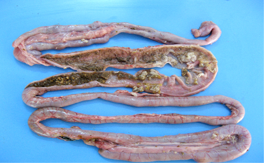
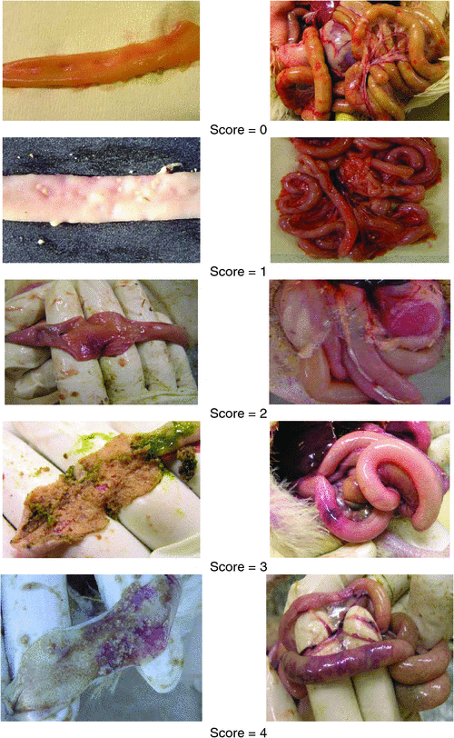
Postmortem lesions :
- SI ( usually middle to distal ) thickened and distended.
- Intestinal mucosa with diphtheritic membrane ( pseudo membrane ) referred to as a “ Turkish Towel”
- Reflux of bile-stained liquid in crop, if upper SI are affected .
- Affected birds tend to be dehydrated and to undergo rapid putrefaction.
- Gall b;ladder distended.,
- Atrophy of spleen, breast muscles and testes.


Microscopic lesions :
- Necrosis of villi of intestinal muscularis.
- Degeneration and necrosis of hepatocytes , proliferation of CT in spleen and bursa.

Diagnosis :
- Clinical signs and lesions
- Demonstration of gram +ve in necrotic lesions in intestine and liver.
DDx :
- Mycoplasmosis
- Respiratory viruses
- Chronic / localized pasteurellosis
- Vitamin A deficiency