Infectious laryngotracheitis ( ILT )
- ILT is an acute , highly contagious infection of chickens and pheasants
- Result in severe production losses due to mortality &/ or decreased egg production
- Severe epizootic form of infection are characterized by signs of respiratory depression , gasping, expectoration of bloody mucus and high mortality
- Mild enzootic form : encountered increasingly in developed poultry industries and manifest variously as mucoid tracheitis, sinusitis, conjunctivitis , general unthriftiness and low mortality
- Distributed world-wide
- The greatest incidence of disease is generally seen in areas of highly intensive poultry production

Route of entry :
- Upper respiratory and ocular routes
Etiology :
Gallid Herpesvirus I , commonly known as infectious laryngotracheitis virus (ILTV)
[ symmetry : icosahedral ]
Transmission :
- It occurs :
- Actively infected birds
- Mechanical transmission ( contaminated equipment and litter )
- No evidence for vertical transmission
- IP = 6-12 days, following natural infection
= 2-4 days , following experimental inoculation
- Morbidity = 50-100%
- Mortality = 10-20%
- Recovered and vaccinated birds are long-term carriers ⬎⬐
Pathogenesis :
Virus
⬐ ⤵
Respiratory tract epithelium conjunctiva
⬇ ⬇
Larynx , trachea , respiratory sinuses swollen watery eyes
air sacs , lungs
⬇
Epithelial damage and hemorrhages
⬇
Nasal discharge , Coughing,
gasping , bloodstained mucus
- Infectious virus usually is present in tracheal tissues and tracheal secretions for 6-8 days PI
- Extratracheal spread of LTV to trigeminal ganglia after 4-7 days of tracheal exposure
- Trigeminal ganglion – principle site of LTV latency
- Reactivation of latent LTV from trigeminal ganglia , 15 months after vaccination of a flock has been reported from Germany
- Apparently spontaneous outbreaks of ILT can occur in field situation
- Rates of shedding of ILTV into trachea could be significantly increased by the stresses of either onset of lay or mixing with unfamiliar birds
⬇
Latently infected chicken can act as unsuspected reservoir host and enable ILTV to infect further susceptible chickens
Clinical signs :
a. Sub – acute form :
- Tracheitis
- Conjunctivitis
- Mild rales
- Nasal and ocular discharge
b. Acute form ( severe epizootic form )
- Nasal discharge
- Moist rales
- Gasping ( pump handle respiration )
- Dyspnea
- Expectoration of blood stained mucus
- Decreased productivity
c. Mild enzootic form :
- Reduced egg production
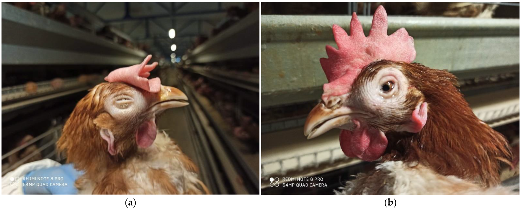
- Watery eyes , conjunctivitis
- Swelling of infra-orbital sinuses
- Mild tracheitis
- Persistent nasal discharge
- Hemorrhagic conjunctivitis

Gross lesions :
- Conjunctiva : edema and congestion
- Infraorbital sinuses : congestion
- Trachea and larynx : blood stained mucus exudates
Mucoid cast ( caseous plug )
- Bronchi : bronchitis
- Air Sacs and lungs : inflammation

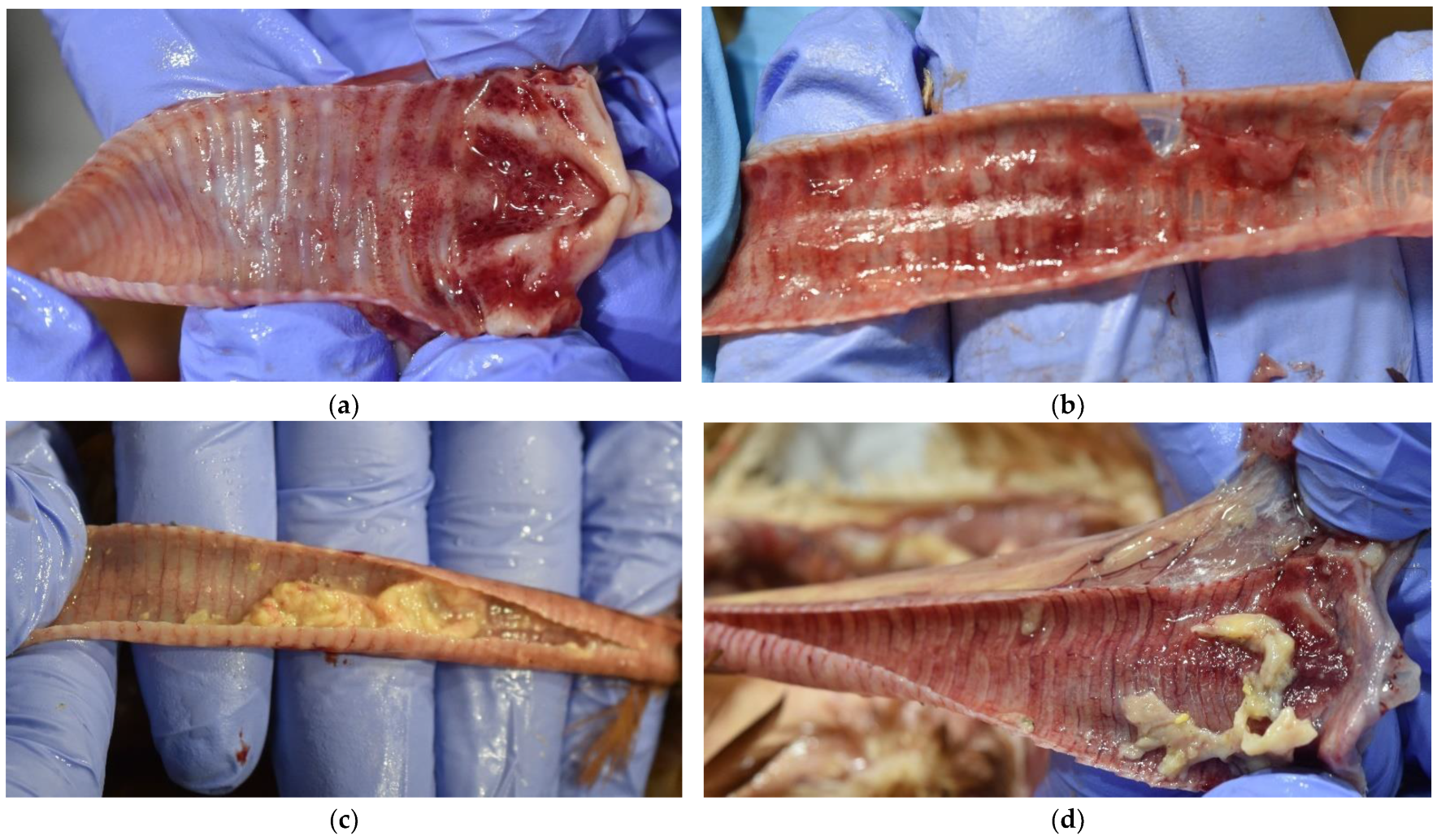
Fig : Severe hemorrhagic tracheitis with mucus due to infectious laryngotracheitis virus in a broiler.
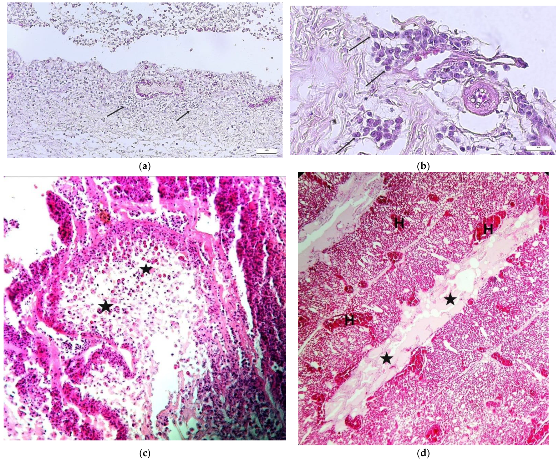
Microscopic lesions :
- Tracheal mucosa : loss of goblet cells
Infiltration of mucosa with inflammatory cells
- Respiratory epithelium : cell enlarge , loss cilia and become edematous
Syncytia are formed
- Lamina propria : blood vessels within lamina propria may protrude into tracheal lumen
- Blood capillaries : rupture of capillaries
- Intranuclear inclusion bodies are found in epithelial cells by 3 days post infection in tracheal epithelial .
Diagnosis :
- History
- Clinical signs and lesions
- Detection of viral antigen
- Histopathology
- Electron microscopy
- FA test and immunoperoxidase test (IPT)
- ELISA
- Detection of viral DNA :
- Dot- blot hybridisation
- PCR
- RT- PCR
- Detection of antibody against LTV :
- Agar gel immunodiffusion (AGID)
- Virus neutralization
- Indirect fluorescent
- Antibody (IFA) test
- ELISA
Fig : ILT. There is fibrinous exudate in the laryngeal opening and also in the tracheal lumen. The tracheal mucosa is hyperemic. A few small plaques of fibrinous exudate are also on the oral and esophageal mucosa