Infectious coryza
- It is usually acute and sometimes chronic, disease of chicken, occasionally pleasant and guinea fowl..
- Characterized by catarrhal inflammation of URT, specially nasal and sinus mucosae.
- It is found worldwide.
- Also known as fowl coryza
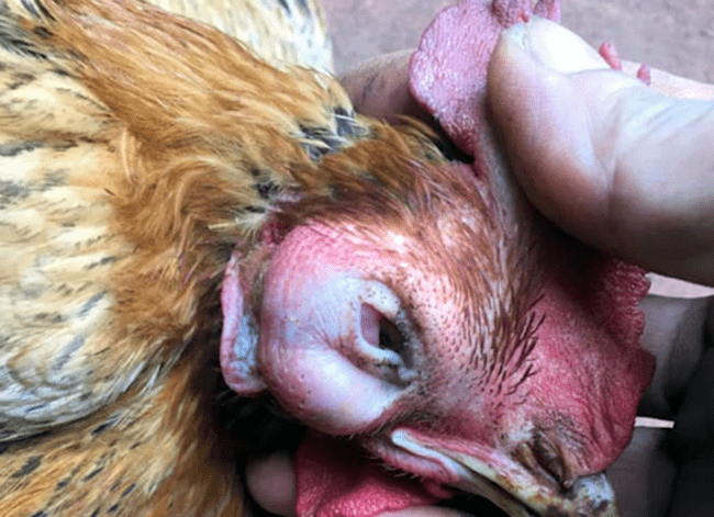
- The disease was named Infectious coryza because it was infectious and affected primarily nasal passage ( nasal discharge, sneezing , swelling of face under eye ).
- All ages are susceptible but occurs most often in adult birds ( layers ) ; so mul;ti- age farms are more susceptible.
- Morbidity is high but mortality is low ( about 20 %)
Etiology :
Avibacterium paragallinarum (Haemophilus paragallinarum )
[gram negative, pleomorphic, non-motile , catalase -ve ;
microaerophilic rod : requires NAD (v-factor) for in vitro growth ]
- Bacteria can survive 2-3 days outside the bird.
Transmission :
- Direct contact , airborne droplets, and contamination of drinking water.
- Egg transmission does not occur.
- Carrier can transmit via exudate and by direct contact
- Ill or sick birds and recovered birds are carrier of disease ( one of the imp source of disease )
- Route of infection :
Conjunctival route , nasal route
Pathogenesis :
Entry of bacteria through route of inhalation
🠋
Bacteria binds to ciliated mucosa of URT
🠋
Bacteria contain capsule and HA which help them to lyse the host cell
🠋
Colonization
🠋
Bacteria release endotoxin
🠋
Pathological lesions
Clinical signs :
- Facial swelling
- Infraorbital sinusitis
- Edema of face around eyes
- Edema of wattle and intermandibular space in male birds
- Purulent ocular and nasal discharge
- Sneezing
- Dyspnoea
- Loss in condition
- Anorexia
- Drop in egg production
- Diarrhoea
- Decrease in growth and development

Postmortem finding :
- In acute; infraorbital sinus ; there is a copious , tenacious , grayish , semifluid exudate .
- Chronic; sinus exudate 🠚 consolidated and turn yellowish
- Other lesions:
- Conjunctivitis
- Eye-lid adherence
- Caseous yellowish material in sinuses
- Tracheitis and bronchitis
- Air sacculitis
- When layers are infected ; there is peritonitis ; due to deposited egg in peritoneal cavity
- Soft shell eggs and egg with hematomas are often seen in ovary

Microscopic :
- Histopathologic response of respiratory organs consists of disintegration and hyperplasia of mucosal and granular epithelia and edema with infiltration of heterophils, macrophages and mast cells.
- Fibrinopurulent cellulitis
- Air sacs: edema thickening, mesothelial hyperplasia , fibrin deposition
Diagnosis :
- History
- Clinical sign and lesions
- Isolation and identification of gram -ve bacteria (bipolar filamentous or coccoid organism )
- Biochemical test :
Catalase test
- PCR
- Serological test : hema agglutination inhibition test ( HI test ) – best one
DDx :
- Mycoplasmosis : chronic respiratory disease
No involvement of infraorbital sinuses ( except turkey )
- Infectious laryngotracheitis : No involvement of infraorbital sinuses
- Newcastle disease : nervous signs
- Fowl cholera : no respiratory signs
- Infectious bronchitis : caseous plug at bifurcation of bronchi
- Vitamin A deficiency