Favus :
- Also known as Avian Ringworm.
- Favus is chronic skin disease that affects poultry and mammals.
- Favus has a worldwide distribution but it’s occurrence has been sporadic .
- Disease is contagious and is transmissible in humans.

Etiology :
Microsporum gallinae ( previously , Trichophyton gallinae )
- Incidence has been reported in chickens, turkey , duck , quail and conary .
- gallinae may be more common in backyard and game chicken flocks .
- Live in damp/ humid places .
Transmission :
- They are transmitted to chickens through direct or indirect contact with skin of other infected birds, animals, insects, people , soil or fomites ( equipment , object , clothes )
Pathogenesis :
Hyphae ( Microsporum gallinae )
⬇
Stratum corneum ( comb, wattle, shanks )
⬇
Hyperplasia and hyperkeratosis
⬇
Infection – confined to epidermis
⬇
Minimal inflammatory reaction
⬇
Dry, white and scaly ( regress , static and progress )
⬇
Feathered areas ( depression around follicles )
[Favus cups ]
Signs :
- White , powdery spots and wrinkled crusts and scab on comb and wattles
- Feather l;oss
- Honeycomb skin
- Thick crusty skin around head
- Loss of condition
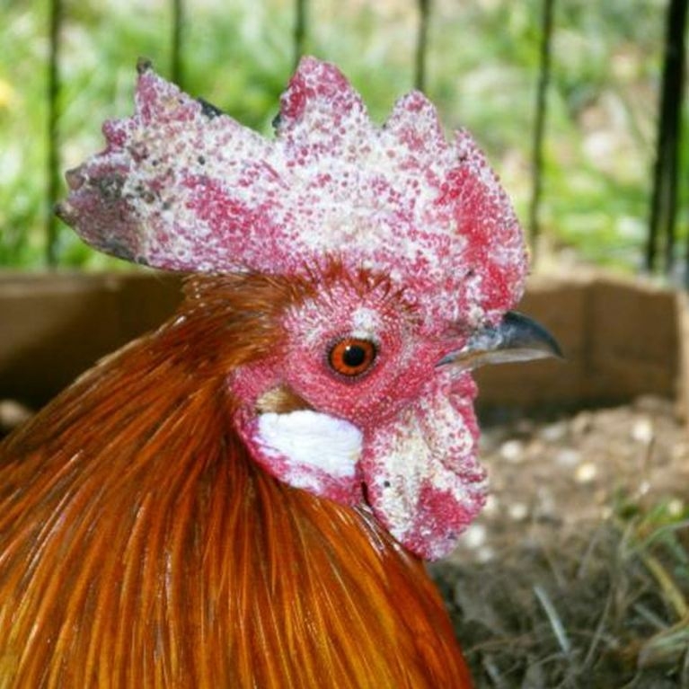

Fig : Microsporum gallinae -positive cock showing white scaling on the comb.
Postmortem lesions :
- Hyperkeratosis of skin epithelium with invasion of stratum corneum by fungal mycelia
- Acanthosis ( brown-to-black, poorly defined, velvety hyperpigmentation of the skin)
- Acantholysis ( is the loss of intercellular connections, such as desmosomes, resulting in loss of cohesion between keratinocytes)
- Hydropic degeneration of cells in stratum spinosum
Microscopic lesions :
- The underlying deris was infiltrated by mononuclear cells and contained lymphoid foci.
Diagnosis :
- History
- Clinical signs
- Physical exams
- Cytology – skin scrapes

Fig : Acanthosis