Coccidiosis :
- It is a parasitic disease of intestinal tract of animals caused by coccidian protozoa
- The disease spreads from one animal to another by contact with infected faeces or ingestion of infected tissue
- Diarrhoea which may become bloody in severe cases is a primary symptoms
- Most animals infected with coccidia are asymptomatic , but young of immunocompromised animals may suffer severe symptoms and death .
Etiology :
- Coccidia are almost universally present in poultry – raising operations , but clinical disease occurs only after ingestion of relatively large no. of sporulated oocyst by susceptible hosts
- Both clinically infected and recovered birds shed oocyst in their dropping which contaminated feed , dust , water , litter and soil .
- Oocyst may be transmitted by mechanical; carriers (eg: equipment , clothing , insects, farms workers and other animals)
- Fresh oocyst are not infective until they sporulate under optimal condition with adequate moisture and oxygen ; this requires 1-2 days
- Coccidia rae host specific and there is no cross-immunity between species of coccidia
- Prepatent period = 4-7 days
- Sporulated oocyst may survive for long periods, depending on environmental factors.
- Oocyst are resistant to some disinfectants commonl;y used around livestock but are killed by freezing or high environmental temperature
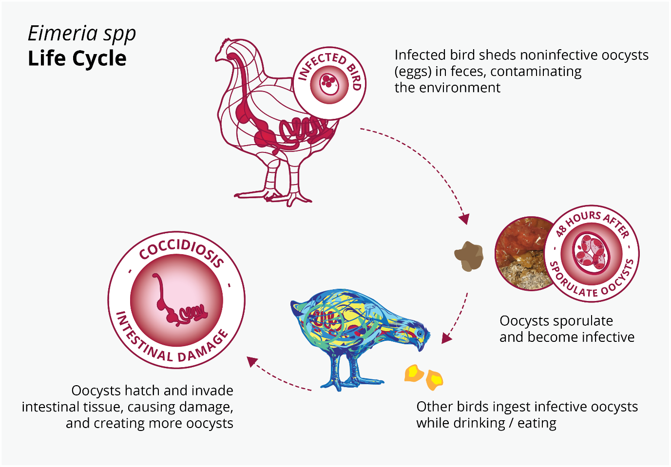
Life Cycle of coccidia :
- The life-cycle is rapid, approximately 4-5 days, and involves massive multiplication from ingestion of a single infective oocyst.
- Both infected and recovered birds shed oocysts. Under conditions of 25-30˚C, this takes 1-2 days.
- Sporulated oocysts have the ability to survive outside the host for very long periods. Since sporulation does not occur below 12˚C or above 39˚C, during winter months spores may remain dormant
- The life-cycle is short and starts with the bird ingesting sporulated oocysts
- The life-cycle is short and starts with the bird ingesting sporulated oocysts
- The sporulated oocysts contain four sporocysts, each containing two sporozoites and the mechanical and acidic environment in the gut result in the release of these sporocysts and sporozoites into the gut.
- The sporozoites invade the duodenal mucosa epithelial cells before undergoing phases of growth and multiplication with periodic release of merozoites into the gut.
- Merozoites develop within the duodenal cells as gametes, in the form of both macro- and microgametocytes
- These develop into a zygote and then an oocyst which is shed in the faeces
- These oocysts require moist conditions to undergo sporulation, a process that requires oxygen and takes about 24 hours, at which point they become infectious.
Pathogenicity :
It is influenced by :
- Host genetics
- Nutritional factors
- Concurrent disease
- Age of host
- Species of coccidia
- Eimeria necatrix and E.tenella are the most pathogenic in chicken . Because schizogony occurs in lamina propria and crypts of SI and caeca , respiratory tract and causes extensive haemorrhage
- Most species develop in epithelial cells lining the villi .
Factor contributing to outbreak :
- Litter moisture content > 30% due to ingress of rain or leaking waterer
- Immunosuppression
- Suboptimal inclusion of anti coccidiosis or incomplete distribution ( poor mixing in feed )
- Environmental and manage mental stress ( overstocking)
- Inoperate feeding system system
- Inadequate ventilation
Clinical signs :
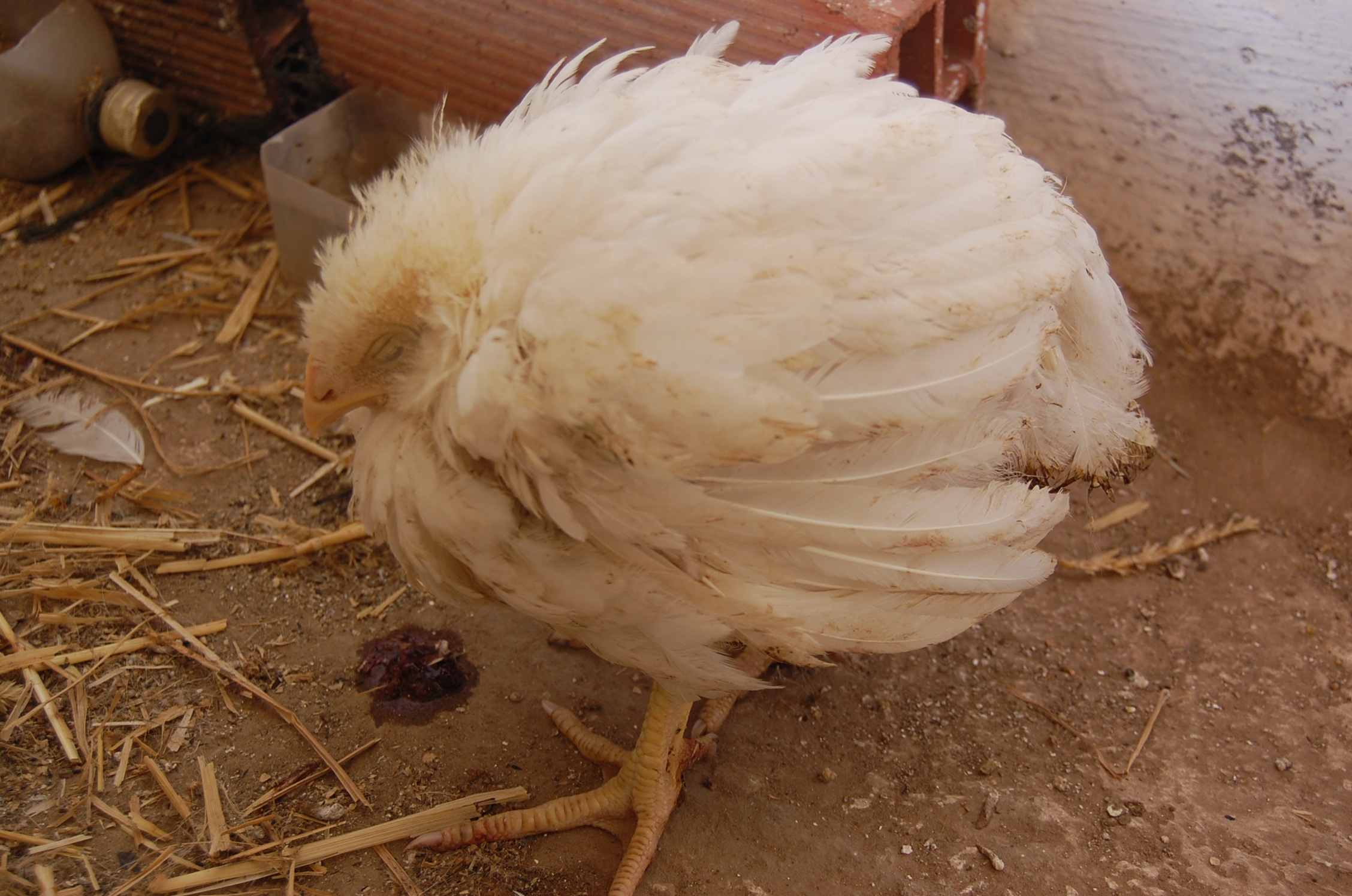
- Sign ranges from decreased growth rate to a high % of visible sick birds
- Severe diarrhoea ( bloody diarrhoea)
- High mortality
- Feed and water consumption are poor
- Weight loss
- Decreased egg production
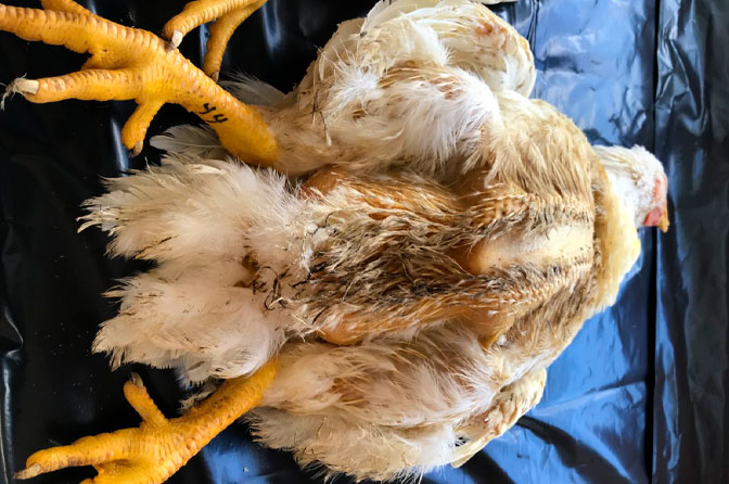
- Ruffled feather
- Dehydration
- Increased anaemia of body and viscera
- Mild infection of intestinal species which would otherwise be classed as subclinical , may cause depigmentation and potentially lead to secondary infection , particularly clostridium spp.

Types :
a. E. tenella :
- They are found only in caeca and can recognized by : accumulation of blood in caeca and by bloody dropping
- Caecal cores , which are accumulation of clotted blood , tissue debris and oocysts , may be found in birds surviving the acute stages.
- Later stage : caecal content becomes thicker , mixed with fibrinous exudate and acquires a cheese-like appearance .
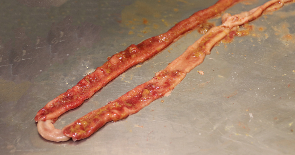
b. E. necatrix :
- Major lesions in anterior and middle portion of SI
- Small white spots , usually intermingled with rounded , bright or dull-red spots of various sizes can be seen on serosal surface
[ this appearance is called – Salt and Pepper]
- In severe cases , intestinal wall is thickened and the infected area dilated to 2-2.5 times the normal
- Lumen may be filled with blood , mucus and fluid
- Fluid loss may result in marked dehydration
- Although the damage is in SI , the sexual phase is completed in caeca
- Oocysts are found only in caeca
- Because of concurrent infection ; oocyst of other species may be found in area of major lesion; misleading to diagnostician
c. E. acervulina
- It is the most common cause of infection
- Lesions include numerous whitish , oval or transverse patches in upper half of SI , which can be easily distinguished on gross examination
- Clinical course in a flock is usually protracted and results in poor growth , an increase in culls and slightly increased mortality
d. E. brunetti :
- It is found in lower SI , rectum, caeca, and cloaca
- In moderate infection , mucosa is pale and disrupted but lacking in discrete foci and maybe thickened
- In severe condition ; coagulative necrosis and sloughing of the mucosa occurs throughout most of SI
e. E. maxima :
- It develops in SI ; where it cause dilation and thickening of the wall
- Petechial haemorrhage
- Reddish , orange or pink viscous mucoid exudate and fluid
- The exterior midgut often has numerous whitish pinpoint foci and the area may appear engorged
- The oocyst and gametocytes ( particularly microgametocytes) , wish are present in lesions are distinctly large
Diagnosis :
- Clinical signs and lesions
- Location in host , appearance of lesions and size of oocyst used in determining specious present
- Confirmed by demonstration of oocyst in faeces or intestinal scrapping
DDx :
- NE (necrotic enteritis)
- UE ( ulcerative enteritis)
- Histomoniasis ( typhlo hepatitis)