Chicken Anaemia Virus (CAV) Infection
[ chicken infectious anaemia, Blue wing disease , Anaemia dermatitis syndrome , Haemorrhagic aplastic anaemia syndrome]
- The disease is characterized by aplastic anemia and generalized lymphoid atrophy with concomitant immunosuppression and frequently is complicated by secondary viral, bacterial, or fungal infections.

Etiology :
- CAV (Chicken anaemia virus ) of Gyrovirus genus of Anelloviridae family
- [ non-enveloped , -ve sense circular DNA ]
Transmission :
- Disease usually during the first 3 weeks of life.

a. Vertical :
- When breeders flocks get infected during the production period ; this occurs especially in younger flocks .
b. Horizontal :
- Through infected organic material or contaminated equipment
- Chickens at any age are susceptible to infection by :
- Oral route
- Respiratory route
- Incubation period :
- CAV is not highly contagious and takes a few weeks to spread through an entire flock.
- IP is relatively long under field condition
- Under experimental infection :
- First signs of anaemia and histologic lesion are seen 8 days after parenteral inocul;ation of virus
- CS generally develop after 10-14 days
- Mortality – 12-14 after inoculation
Pathogenesis :
Virus ➦ oral / respiratory route
⬇
Principle sites of replication are hemocytoblasts in bone marrow , precursor T cells in the cortex of thymus , dividing Tcd4 and Tcd8 cells in the spleen .
Replication and destruction of hemocytoblasts in bone marrow
⬇
Decrease production of RBC and WBC
⬇
Anaemia
Replication and destruction of T-cells ➦ immunosuppression
- CAV can be found latent in SPF flocks where viruses are detected in ovaries , oviduct , testicles , and spleen of birds without obvious seroconversion until the birds come into production .
Clinical signs :
- Anaemia ( PCV ≤ 27)
- Paleness
- Anorexia
- Lethargy
- Depression
- Watery and slowly clotting bird
- Reduced weight gain
- High mortality rates resulting from secondary complicating infections ( generally 10-20% , but as much as 60% )
- Anaemia , leukopenia and pancytopenia seen often on blood smears
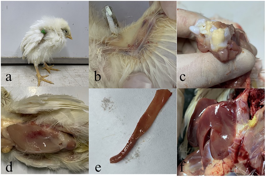
Gross :
- Organs are pale
- Thymus is generally atrophied ( sometimes absence of thymic lobes )
- Bursa of fabricius may be small .
- Bone marrow is pale or yellow
- Hemorrhages may be present in or under skin and in muscles and other organs
- Lesions associated with secondary infections may be present
- Hemorrhages in proventricular mucosa and subcutaneous and muscular hemorrhages are sometimes associated with severe anaemia
- Gangrenous dermatitis on feet , legs wings or neck
- Acute ,mycotic pneumonia
- Discoloured liver and kidney

- Focal lesions ( wings ) appear as ecchymotic skin hemorrhages
- Lesions turn blue and may break, releasing serosanguineous exudate which is prone to secondary bacterial infections leading to gangrenous dermatitis
- This can be especially notorious at end of wings ; hence named “Blue wing disease “
- Tips of wings may appear hemorrhagic and necrotic
Microscopic lesion :
- Lymphoid cell population are depleted in primary primary and secondary lymphoid organs
- Severe lymphocytic depletion in thymic cortex
- Granulocytic and erythrocytic compartments in bone marrow are atrophied or hypoplastic .

Diagnosis :
- Clinical signs
- Gross lesion
- Demonstration of seroconversion in parent flock
- Isolation and identification of virus in lymphoblastoid cell line
- Serology