Avian encephalomyelitis :
- Avian encephalomyelitis (AE) is a viral disease of the CNS of young chickens, turkeys, Japanese quail, pheasants, and pigeons.
- Turkeys are less susceptible to natural infection and generally develop a milder clinical disease than chickens.
- Ducklings and guinea fowl are susceptible to experimental infection.
- AE is characterized by neurologic signs that result from infection of the CNS with an RNA virus.
Etiology :
AEV ( Tremovirus A in the genus Tremovirus of the family Picornaviridae)
- Disease is most common in chickens (1-6 weeks of age)
- IP = varies from 5-14 days depending upon route of infection
- Pathotype ( two) :
- Enterotropic – natural field strains
- Embryo -adapted strains
Transmission :
- Egg transmission is major route of transmission
- Horizontal transmission by direct contact (more common)
- Natural field strains of the virus are enterotropic and enter into the body via oral route and multiply in the intestine.
- Infected birds shed the virus in their feces for a few days to a few weeks, which serves to spread the infection to hatchmates.
- AE virus is resistant to environmental conditions and may remain infectious for long periods.
Clinical signs :
a. Vertically infected chicks :
- Clinical signs appear during the first week after hatching, although signs may be present in a few birds at hatching.
- Vertical infection followed by horizontal infection causes a characteristic biphasic mortality pattern.
b. Horizontally infected :
- Infected chicks usually show clinical signs at 2–4 weeks of age
Common signs:
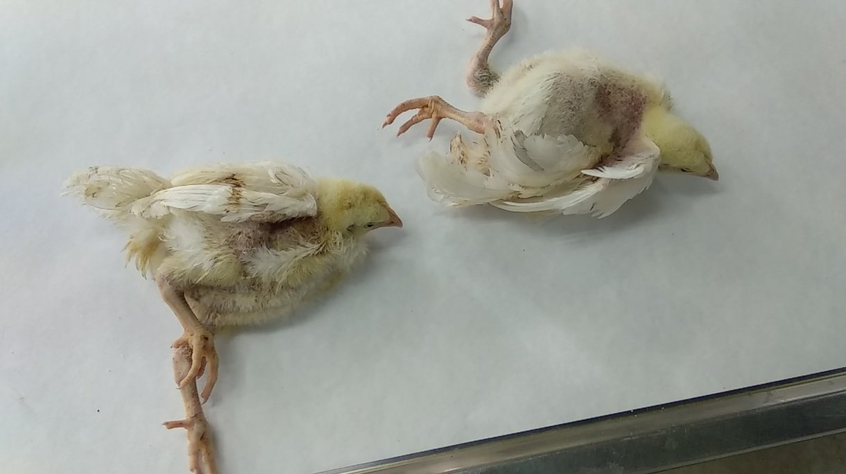
- Ataxia
- Tremors
- Weakness that progresses to paralysis and recumbency
- Nervous sign
- Dull expression of the eyes, followed by unsteadiness,
- Sitting on hocks,
- Tremors of the head and neck,
- Paresis (weakness or partial paralysis), and finally total paralysis.
- Feed and water consumption decreases
- Birds will lose weight.
- Drop in egg production
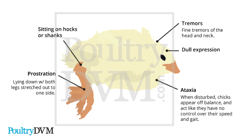
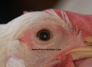
fig : A three-week-old pheasant poult showing cataract in the eye
Postmortem lesions
Gross lesion :
- No gross lesions are seen in the brain of birds infected with avian encephalomyelitis.
- Gray to white foci may be visible on cut surfaces of the muscle of the gizzard.
- Weeks after infection, opacity of eye lenses (cataracts) may occur in a small percentage of chickens that survive the infection.

Microscopic :
- Lesions in the CNS are found in the brain (cerebral peduncle, cerebellum, brain stem) and spinal cord and consist of degeneration and necrosis of neurons, perivascular lymphocytic cuffing, and gliosis with formation of glial nodules.
- In the cerebellum, there are areas of necrosis or loss of Purkinje cells and replacement by glial nodules that extend into the molecular layer of the gray matter.
- Neuronal lesions of central chromatolysis, shrinkage and increased basophilia, satellitosis, and neuronophagia are best found in neuron clusters (nuclei) in the brain stem, arbor vitae of the cerebellum, and lateral horn (gray matter) of the spinal cord.
- Outside the CNS is diffuse or nodular lymphocytic infiltrates in the gizzard muscle, muscular layer of the esophagus and proventriculus, myocardium, and pancreas

Diagnosis :
- History
- Clinical signs
- Characteristics histopathological lesions in brain and spinal cord
- Detection of viral antigen :
- Virus neutralization test
- Agar gel test
- Embryo susceptibility test
- Isolation and identification of virus
Differential diagnosis :
- Bacterial or mycotic encephalitis
- Rickets and nutritional encephalomalacia
- Newcastle disease
- Vit (E,A, riboflavin ) deficiency
- Toxicities
- Marek’s disease
- Brain abscess
- Enterococcus hirae infection
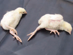
Fig: The infectious encephalomyelitis (IEM) is characterized by signs of ataxia, progressing to paralysis.The tremor could be unapparent, but is often perceptible when the chicken is held gently with the hand and carefully looked at. the expression of the eye is dull.
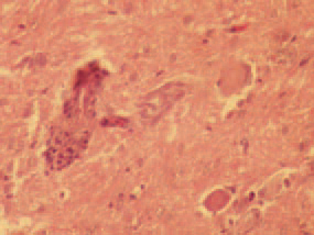
fig : The histological lesions are specific and with a diagnostic value. A nonpurulent encephalomyelitis with marked perivascular clusters is present.
Fig: A particularly valuable finding is the central chromatolysis (A) of neurons in segments from the lumbosacral widening of the spinal cord and more rarely, chromatic pyknosis
fig: Valuable diagnostic findings are the dense lymphoid clusters in the muscles of the proventriculus and the gizzard (A) and in the pancreatic interstitium (B).